
Executive Overview
For decades, the opening chapters of vertebrate and invertebrate life were written under a pervasive scientific assumption: that the DNA within a newly fertilized egg was a structural "blank slate." Conventional wisdom dictated that the maternal and paternal genomes initially coalesced as a loose, disorganized, and chaotic tangle of chromatin. According to this long-standing dogma, the genome remained a structural amorphous mass until the embryo achieved a critical developmental milestone known as Zygotic Genome Activation (ZGA)—the moment the embryonic genome officially "woke up" and began transcribing its own distinct genetic instructions.
That foundational paradigm has now been shattered.
In a pair of landmark studies published in Nature Genetics and Nature Cell Biology, an international consortium of researchers has unveiled a vastly different reality. Spearheaded by Professor Juanma Vaquerizas and his team, alongside collaborators like Professor Ulrike Kutay at ETH Zürich in Switzerland, scientists have demonstrated that the genome exhibits an astonishing, highly regulated level of three-dimensional architecture long before ZGA ever occurs.
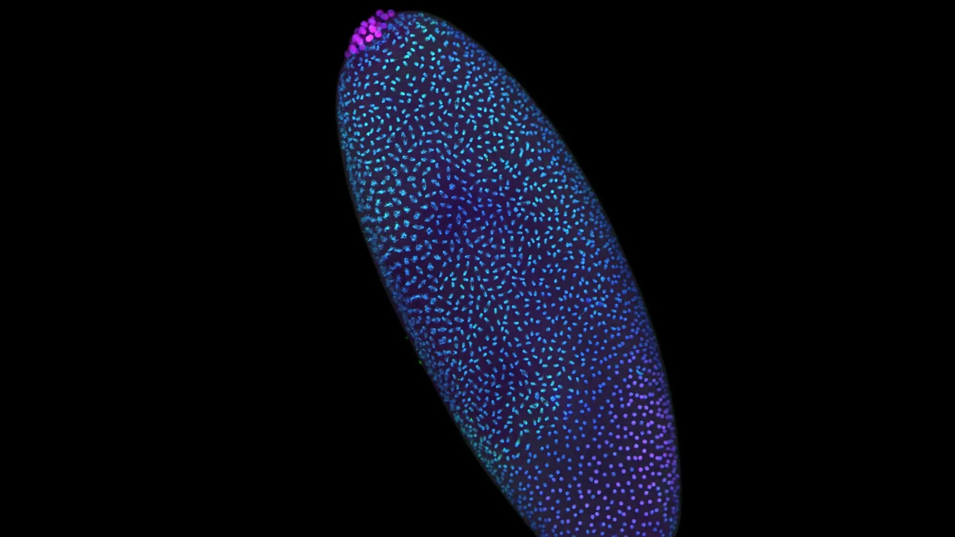
To achieve this breakthrough, the research team engineered a pioneering molecular technique known as Pico-C. This ultra-sensitive technology allows researchers to map the 3D folding of DNA with unprecedented spatial resolution while requiring a fraction of the biological material previously demanded by standard genomic assays. By deploying Pico-C on fruit fly (Drosophila) embryos, the team discovered that an elaborate, modular structural scaffold is meticulously erected hours before the genetic "on" switch is ever flipped.
The implications of this discovery reach far beyond basic embryology. In human cells, the researchers demonstrated that when the molecular anchors responsible for maintaining this delicate 3D architecture are compromised, the consequences are catastrophic. Cells mistake the structural collapse for an active viral invasion, triggering a false innate immune response that can spark destructive inflammation and disease. By linking the precise construction of life at its inception with the devastating systemic effects of its deterioration, these findings bridge a critical gap in our understanding of genetics, cellular biology, and human pathology.
Detailed Chronology: From Biological Chaos to Precision Architecture
To appreciate the magnitude of the recent discoveries, it is necessary to retrace the timeline of how geneticists historically viewed the earliest moments of embryonic development, and how the newly developed Pico-C technology fundamentally shifted that trajectory.
The Traditional Paradigm: The "Blank Slate" Hypothesis
Historically, textbook biology described fertilization as a violent yet miraculous reset. When a sperm fertilizes an oocyte, the two distinct pronucleus sets merge, bringing together two separate genetic packages. For years, scientists observed that the early embryonic cell cycles—characterized by rapid, synchronous divisions without intervening growth phases—were transcriptionally silent.
Because the embryo was not yet actively producing its own messenger RNA (mRNA) and relied entirely on maternal stockpiles laid down during oogenesis, researchers inferred that the underlying chromatin must be unstructured. The prevailing hypothesis characterized the pre-activation genome as a passive, fluid state of structural chaos. The standard view assumed that genome folding was a consequence of transcription—meaning that genes only organized themselves spatially after they were awakened and began expressing proteins.
The Turning Point: The Development of Pico-C
This model persisted largely due to technical limitations. Standard chromosome conformation capture methods, such as Hi-C, required millions of cells to construct a reliable map of DNA interactions. Because early embryos are scarce and precious, obtaining sufficient material to analyze minute-by-minute architectural changes was virtually impossible.
Enter Pico-C, a breakthrough technology engineered specifically to overcome these sample-size limitations. Pico-C operates with approximately ten times less input material than conventional techniques, opening an unprecedented window into rare cell populations and early embryonic stages.
By applying Pico-C to rapidly dividing fruit fly embryos, the research team tracked the exact physical rearrangements of chromatin during the first few hours post-fertilization. The results overturned decades of assumptions. Instead of a disorganized soup of DNA, the researchers observed a highly choreographed construction project. Long before Zygotic Genome Activation, chromosomes began folding into precise loops, domains, and modular compartments. This spatial arrangement systematically positioned regulatory elements—such as enhancers—in close physical proximity to their target genes, priming the genome for action long before a single gene was transcribed.
The Human Cell Translation: Architecture and Catastrophe
Once the blueprint of early architectural construction was mapped in model organisms, the research consortium turned its attention to human somatic and embryonic cells. In a companion study published in Nature Cell Biology, Professor Ulrike Kutay and her team at ETH Zürich investigated what happens when this intricate architecture fails.
Using high-resolution mapping strategies akin to Pico-C, the researchers systematically disrupted the molecular "anchors"—specific proteins and structural complexes responsible for stabilizing the genome’s 3D folding. When these anchors were experimentally removed, the spatial organization of the chromatin collapsed.
The cellular response to this structural failure was swift and dramatic. Human cells possess ancient, highly sensitive surveillance systems designed to detect foreign nucleic acids, such as viral DNA invading the nucleus. When the host genome’s spatial architecture fell apart, the nuclear environment changed in ways that the cell’s surveillance machinery misinterpreted as a viral takeover. The cell initiated an innate immune response, mounting defenses against its own genetic material. This false alarm cascades into chronic inflammation, cellular dysfunction, and, ultimately, severe pathological states.
Supporting Context, Methodology, and Metrics
The rigorous execution of these studies required cutting-edge experimental design, innovative molecular biology tools, and meticulous data analysis. Below is a breakdown of the organisms, technologies, and quantitative frameworks that underpinned the research.
The Model Organism: Drosophila melanogaster
The fruit fly remains a cornerstone of genetic discovery for several reasons that made it the ideal proving ground for Pico-C:
- Rapid Embryogenesis: Following fertilization, the fruit fly embryo undergoes rapid nuclear divisions within a shared cytoplasm (syncytial blastoderm stage), producing thousands of nuclei in a matter of hours.
- Synchronized Development: The predictability and speed of these early divisions allow researchers to isolate precise developmental windows with high temporal accuracy.
- Genetic Conservatism: Despite its evolutionary distance from humans, the fundamental mechanisms governing chromatin folding, transcriptional regulation, and nuclear architecture are remarkably conserved across species.
Quantitative Breakdown of Pico-C Performance Metrics
| Feature / Metric | Standard Hi-C Technology | Pico-C Technology | Biological Significance |
|---|---|---|---|
| Minimum Sample Input | Millions of cells ($10^6$ range) | ~10 times less material ($10^5$ or fewer cells) | Enables spatial genomic analysis of rare early embryos and scarce clinical samples. |
| Spatial Resolution | Moderate to High (dependent on depth) | Ultra-High spatial fidelity | Detects subtle, localized DNA looping events prior to gene activation. |
| Temporal Precision | Broad developmental windows | Minute-by-minute developmental tracking | Captures the exact sequence of architectural scaffolding erection. |
| Artifact Reduction | Prone to ligation noise in low-input settings | Optimized molecular capture protocols | Delivers cleaner contact matrices with reduced background noise. |
The Mechanics of Chromatin Folding and Gene Regulation
To understand why this early 3D scaffolding matters, one must examine how the linear strand of DNA operates inside the microscopic confines of a cell nucleus. Stretched out, a human DNA molecule measures approximately two meters in length, yet it must fit inside a nucleus just micrometers wide.
This packaging is not random. DNA wraps around histone proteins to form chromatin, which then folds into loops, topologically associating domains (TADs), and larger compartments. These folds bring distant genomic regions into physical contact. An enhancer sequence—located thousands of base pairs away from a gene—can stimulate that gene’s transcription only if the 3D folding of the DNA bends the intervening space, allowing the enhancer to physically touch the gene’s promoter.
By demonstrating that this folding occurs before transcription begins, the research proves that the genome’s physical architecture acts as a pre-set switchboard. The wires are plugged in and organized before the power is ever turned on.
Official Statements and Expert Insights
The collaborative nature of these studies brought together experts in computational biology, structural genomics, and cellular signaling. Their insights illuminate the profound philosophical and practical shifts happening within modern molecular biology.
Professor Juanma Vaquerizas, senior author of the Nature Genetics study, reflected on the psychological shift within his research group as the data emerged:
"We used to think of the time before the genome awakens as a period of chaos. But by zooming in closer than ever before, we can see that it’s actually a highly disciplined construction site. The scaffolding of the genome is being erected in a precise, modular way, long before the ‘on’ switch is fully flipped."
Noura Maziak, lead author of the study, emphasized the technological leap that made these discoveries possible:
"Traditional methods forced us to look at development through a blurry lens because we simply needed too much biological material to get a clear picture. With Pico-C, we can now observe individual developmental milestones with a level of clarity that transforms our understanding of how life initiates its program."
Connecting the two halves of the research—the construction of life versus the consequences of structural collapse—Professor Vaquerizas synthesized the overarching narrative of the twin publications:
"These two studies tell a complete story. The first shows us how the genome’s 3D structure is carefully built at the start of life. The second shows us the disastrous consequences for human health if that structure is allowed to collapse."
Professor Ulrike Kutay, co-author of the companion human cell study at ETH Zürich, highlighted the translational medical implications of the findings:
"When the molecular anchors that maintain nuclear architecture are lost, the cell does not merely experience a structural defect; it undergoes an identity crisis. By misinterpreting its own unfolded DNA as a viral pathogen, the cell triggers an immune response against itself. This provides a direct mechanistic link between nuclear disorganization and inflammatory pathologies in humans."
Future Outlook: Implications for Medicine, Developmental Biology, and Beyond
The revelation that the genome is pre-patterned structurally before transcriptional activation opens an expansive new frontier in biomedical research. As laboratories around the world begin adopting Pico-C and similar high-sensitivity mapping tools, several key areas of research are poised for transformation.
1. Redefining Developmental Disorders and Congenital Defects
Many developmental abnormalities originate during the earliest hours following fertilization, long before a mother might even know she is pregnant. If the initial scaffolding of the genome is erected incorrectly—due to genetic mutations, environmental toxins, or metabolic stress—the downstream consequences can be devastating. Understanding the precise choreography of early DNA folding provides a new diagnostic framework for investigating congenital disorders, miscarriages, and unexplained developmental arrests.
2. Novel Insights into Autoimmune and Inflammatory Diseases
The discovery that structural collapse of chromatin triggers an innate immune response opens exciting avenues in immunology. Researchers can now investigate whether chronic inflammatory conditions, premature aging syndromes (such as progeria), and certain autoimmune disorders stem from subtle, progressive failures of nuclear architecture and anchoring proteins. By identifying the specific molecular anchors that stabilize genome folding, pharmacologists may one day develop targeted therapies designed to reinforce nuclear structure and prevent false immune alarms.
3. Advancements in Regenerative Medicine and Stem Cell Reprogramming
In regenerative medicine, scientists routinely reprogram somatic cells back into pluripotent stem cells (iPSCs) by introducing specific transcription factors. These reprogramming protocols essentially force a mature cell to wipe its developmental slate clean and revert to an embryonic-like state. Understanding how genomes naturally construct their 3D scaffolding at the dawn of life will provide bioengineers with a master blueprint, allowing them to optimize reprogramming protocols, enhance cellular fidelity, and generate safer, more stable stem cell lines for therapeutic applications.
4. Technical Refinement and Single-Cell Genomics
While Pico-C represents a massive leap forward by reducing sample requirements tenfold, the relentless march of genomic technology points toward single-cell 3D genomics. Future iterations of these technologies will allow scientists to trace the 3D folding of individual embryonic cells in real-time as they divide and differentiate. This granular tracking will resolve long-standing debates regarding cellular heterogeneity during early development.
Funding and Acknowledgments
This groundbreaking research was supported by prestigious grants from the Medical Research Council (MRC) and the Academy of Medical Sciences (AMS) through an AMS Professorship award, ensuring that the investigative team could push the boundaries of spatial genomics and uncover the hidden architecture that governs the very dawn of life.