
Executive Overview
Deep within the structural architecture of the modern human brain sits the pineal gland—a tiny, pinecone-shaped endocrine structure historically dubbed by philosopher René Descartes as the "principal seat of the soul." For centuries, science has understood its role in regulating our circadian rhythms through the secretion of melatonin, modulating sleep-wake cycles in response to environmental light. However, groundbreaking evolutionary research has just upended our understanding of how this enigmatic gland came to be.
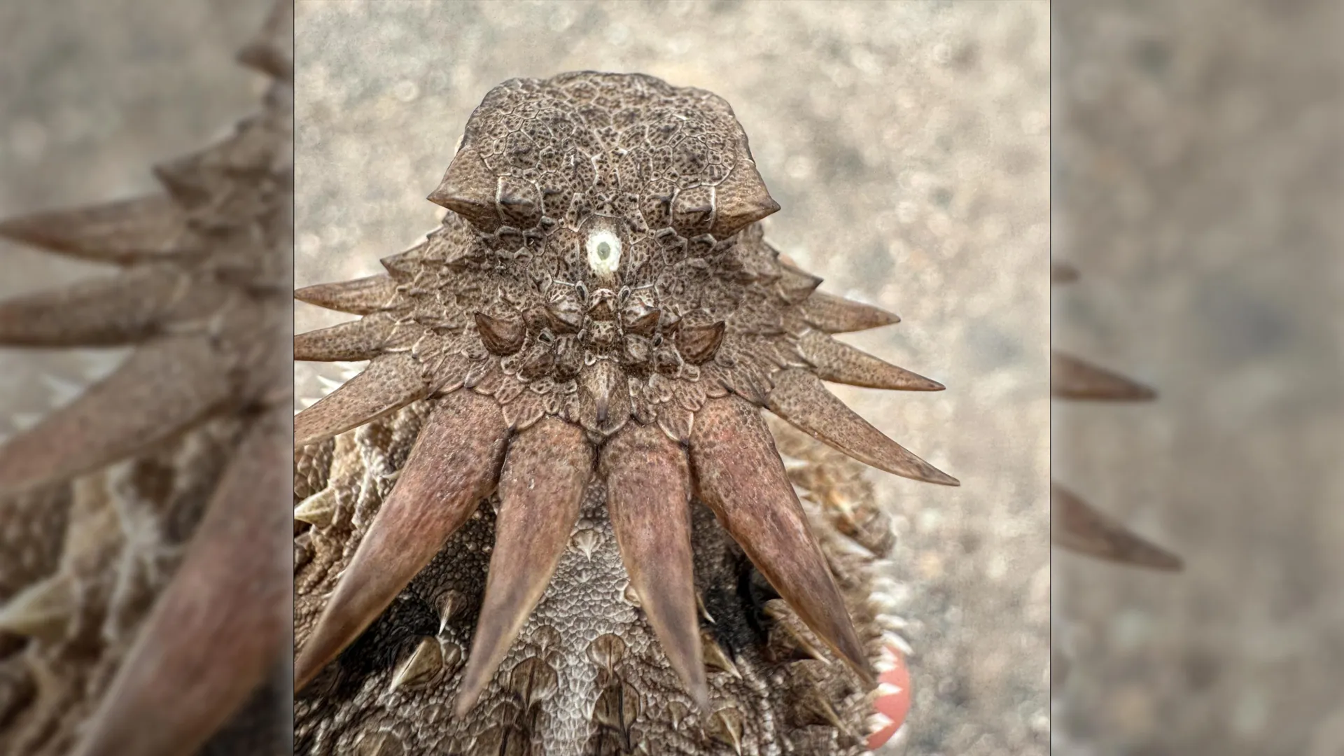
According to a collaborative study published by evolutionary biologists from Lund University in Sweden and the University of Sussex in the United Kingdom, all living vertebrates—from mice and fish to birds, reptiles, and humans—trace their lineage back to a bizarre, worm-like organism that lived nearly 600 million years ago. This distant evolutionary relative possessed a single, central light-sensing organ positioned squarely on top of its head, evoking the mythological archetype of a tiny cyclops.
This paradigm-shifting research does more than merely add a strange footnote to the history of life on Earth; it fundamentally rewires our understanding of neurobiology and sensory evolution. The findings demonstrate that the human pineal gland is not an evolutionary novelty, but rather the direct, repurposed remnant of a primordial median eye. Furthermore, this discovery resolves a long-standing evolutionary mystery: why vertebrate eyes—including our own—are fundamentally constructed in a manner radically different from the eyes of invertebrates like insects, octopuses, and squids.
By detailing an extraordinary evolutionary detour—in which our ancestors lost their original paired eyes, survived via a single central photosensitive spot, and subsequently rebuilt an entirely new visual system from the brain outward—this study provides unprecedented insight into the deep history of human biology.
Detailed Chronology: The Evolutionary Trajectory of Vertebrate Vision
To comprehend how humanity inherited its neurological architecture from a microscopic, plankton-feeding worm, scientists must look back into the Ediacaran and early Cambrian periods, an epoch of explosive biological innovation and environmental transformation.
Phase I: The Ediacaran Divergence and the Stationary Life
Approximately 600 million years ago, the Earth’s oceans played host to a myriad of soft-bodied, enigmatic organisms that bore little resemblance to the fauna dominating modern marine ecosystems. Among them was a small, elongated, worm-like creature that anchored itself to the seafloor or sediment, adopting a sessile, stationary lifestyle.
To survive without the ability to actively hunt or flee, this organism relied on passive feeding mechanisms, filtering microscopic plankton and organic detritus from the passing seawater. Deep phylogenetic analysis suggests that even earlier in its lineage, this creature likely possessed paired, lateral eyes—a common trait among early bilaterally symmetrical animals. These rudimentary visual structures likely helped ancestral species navigate open waters or detect passing shadows.
However, evolution is brutally efficient, discarding biological machinery that no longer serves an organism’s immediate survival imperatives. As the worm-like ancestor settled into its sedentary, bottom-dwelling niche, maintaining complex paired eyes became energetically costly and functionally obsolete. Over countless generations, these lateral visual organs degenerated and disappeared entirely.
Phase II: The Rise of the Median Cyclops
Though the paired eyes were lost, the ancient organism did not become entirely blind to its environment. A cluster of light-sensitive cells persisted in the exact center of its head. Protected by surrounding tissue and connected to early neural clusters, these photosensitive cells gradually coalesced into a functional, singular "median eye."
This median eye was not capable of forming complex, high-resolution images of the surrounding world. Instead, it served as an ecological compass and survival alarm. By detecting the cyclical transitions between light and darkness, the median eye allowed the organism to sense orientation, track the time of day, and respond to the passing shadows of potential predators overhead. For millions of years, this single-eyed configuration served as the primary sensory window through which our lineage interacted with light.
Phase III: The Return to the Pelagic Zone and the Rebuilding of Vision
The evolutionary trajectory of this lineage shifted dramatically when descendants of this sessile worm abandoned their stationary lifestyle and returned to an active, swimming existence in the pelagic zone. This ecological transition placed immense selective pressure back onto the organism’s sensory systems. Navigating a dynamic, three-dimensional aquatic environment required sophisticated spatial awareness, obstacle avoidance, and target tracking—capabilities that a simple light-detecting median spot could not provide.
Rather than resurrecting the long-lost lateral eyes of its more distant ancestors, evolution improvised a radically innovative solution. Portions of the original median eye system, intimately connected to the developing central nervous system, began to expand and differentiate. Through a series of complex genetic and developmental shifts, these neural tissues gave rise to entirely new, paired image-forming eyes.
This unexpected sequence of events—the loss of ancestral paired eyes, a prolonged phase relying solely on a median cyclopean eye, and the subsequent de novo construction of modern eyes from neural tissue—explains the unique structural anatomy that defines all vertebrate vision today.
Supporting Context & Metrics: The Architectural Divide in Animal Vision
For over a century, comparative anatomists have marveled at the profound structural differences between the eyes of vertebrates and those of successful invertebrate lineages, such as arthropods (insects, crustaceans) and mollusks (squids, octopuses). Until now, the developmental origins of these disparities remained stubbornly opaque.
The Inside-Out Vertebrate Retina
In humans and all other vertebrates, the eye develops as an direct extension of the embryonic brain. During embryogenesis, the optic vesicles push outward from the diencephalon toward the surface ectoderm. Consequently, the vertebrate retina is structurally "inverted"—meaning that light must pass through layers of ganglion cells, amacrine cells, bipolar cells, and blood vessels before finally striking the light-sensitive photoreceptor cells (rods and cones) nestled at the very back of the retina.
While this anatomical arrangement seems counterintuitive from an engineering perspective, the new Lund-Sussex research clarifies why it evolved this way: because the foundational sensory cells originated from within the central nervous system itself, rather than from external integumentary tissues.
The Inverted Blueprint of Invertebrate Eyes
Conversely, insects, spiders, and cephalopods like squids and octopuses construct their visual systems via an entirely different developmental pathway. Their eyes originate directly from the surface ectoderm—the skin on the sides of the head. As a result, the photoreceptor cells in an insect’s compound eye or a squid’s camera-type eye point directly toward the incoming light source, with their axons projecting backward into the optic lobes of the brain.
The discovery of the 600-million-year-old cyclopean ancestor bridges this historical divide. It demonstrates that vertebrate vision took an entirely separate evolutionary track, bypassing the skin-derived visual origins shared by most other phyla in favor of a brain-centrism forged during the era of the median eye.
Official Statements and Expert Analysis
The implications of this study are reverberating through the international scientific community, challenging entrenched textbook models of neuro-evolutionary history.
"The results are a surprise. They turn our understanding of the evolution of the eye and the brain upside down," notes Dan-E Nilsson, professor emeritus in sensory biology at Lund University and one of the lead authors of the research.
Prof. Nilsson emphasizes that tracing the structural lineages of photosensitive proteins and neural circuits required an unprecedented synthesis of comparative genomics, developmental biology, and fossil-record analysis.
"For the first time, we now also understand the origin of the neural circuits that analyze the image in our retina," Nilsson explains. By establishing that the foundational neural wiring of the vertebrate visual system evolved to process signals from a central median organ before branching into paired visual structures, researchers can now map out the step-by-step genetic scaffolding that allowed complex brains to interpret visual data.
The transition from a primary light-sensing spot to an endocrine regulator of physiology is equally staggering.
"It’s mind-boggling that our pineal gland’s ability to regulate our sleep according to light stems from the cyclopean median eye of a distant ancestor 600 million years ago," Nilsson concludes.
Other evolutionary neurobiologists unaffiliated with the study have praised the rigorous methodology, noting that the integration of deep-time comparative anatomy with modern molecular genetics provides a robust framework that resolves decades of debate regarding vertebrate eye origins.
Future Outlook and Clinical Implications
As evolutionary biologists continue to refine our understanding of the Cambrian and Ediacaran transitions, the ripple effects of this discovery extend far beyond theoretical phylogenetics. Understanding the deep evolutionary history of the pineal gland and the vertebrate retina holds profound potential for modern biomedicine, neuropharmacology, and regenerative medicine.
Decoding Circadian Disorders and Sleep Medicine
The pineal gland remains the master conductor of human chronobiology. By synthesizing melatonin in response to suprachiasmatic nucleus signals driven by retinal exposure to sunlight, it dictates our circadian rhythms. Modern society—marred by chronic artificial light exposure at night, shift work, and ubiquitous blue-light-emitting screens—frequently experiences circadian desynchronization, which is increasingly linked to metabolic disorders, cardiovascular disease, depression, and neurodegeneration.
Understanding that the pineal gland’s photosensitive origins date back 600 million years highlights how deeply embedded these biological rhythms are within our genomic heritage. Researchers suggest that studying the ancient, conserved genetic pathways governing pineal development and function could inspire novel therapeutic interventions for sleep disorders and seasonal affective disorder (SAD).
Advancing Retinal Therapeutics and Neuro-Engineering
Furthermore, recognizing the retina as an direct structural and developmental extension of the brain provides fresh perspectives for neuro-ophthalmology. Conditions such as retinitis pigmentosa, macular degeneration, and traumatic optic neuropathy involve the degradation of neural tissue that shares an ancient lineage with the central nervous system.
By mapping the genetic instructions that allowed our ancestors to rebuild an image-forming visual system from brain tissue, bioengineers and stem-cell researchers gain valuable blueprints for retinal tissue engineering. Regenerative therapies aimed at growing replacement retinal cells or interfacing electronic bionic eyes directly with the visual cortex stand to benefit immensely from a comprehensive understanding of how these neural circuits were originally assembled over deep evolutionary time.
Conclusion
Ultimately, this research serves as a humbling reminder of humanity’s deep, intricate connection to the history of life on Earth. When humans look up at the night sky, adjust to a changing time zone, or drift off to sleep under the influence of melatonin, they are utilizing biological machinery forged in the murky, ancient seas of a pre-Cambrian world. The tiny cyclops that once rested atop the head of a stationary, worm-like ancestor lives on quietly within us all—a testament to evolution’s remarkable capacity to repurpose, rebuild, and innovate across hundreds of millions of years.