
EXECUTIVE SUMMARY
For centuries, evolutionary biologists have mapped the complex architecture of the vertebrate eye with a sense of architectural wonder. From the eagle soaring high above mountain peaks to the human eye reading these very words, our visual apparatus is a masterpiece of biological engineering. Yet, beneath its intricate design lies an evolutionary oddity that has baffled scientists for generations: why are vertebrate eyes fundamentally inverted compared to those of invertebrates like squid and insects?
Groundbreaking new research spearheaded by sensory biologists at Lund University and the University of Sussex has blown the lid off this long-standing anatomical mystery. According to their findings, every living vertebrate—including humans, birds, reptiles, amphibians, and fish—shares an astonishingly strange ancestor that passed through a distinctly "cyclops-like" stage nearly 600 million years ago.
This prehistoric organism, a tiny, worm-like creature drifting in the ancient Ediacaran oceans, abandoned its original paired eyes when it adopted a stationary, filter-feeding lifestyle. In their place, a single, central median eye emerged on the top of its head. Millennia later, when its descendants returned to an active, swimming lifestyle, evolution performed a stunning pivot: it repurposed portions of this single central eye to construct an entirely new pair of image-forming eyes.
This evolutionary detour not only solves the riddle of why vertebrate retinas develop outward from the brain rather than inward from the skin—unlike the camera-like eyes of cephalopods and arthropods—but it also reveals a ghost in our biological machine. According to the research team, led by Professor Emeritus Dan-E Nilsson, the evolutionary remnants of this ancient median eye survive to this day, deeply embedded within the modern human brain as the pineal gland, the master regulator of our sleep-wake cycles.
The implications of this study reach far beyond comparative anatomy, turning foundational assumptions about the evolution of visual and neural systems completely upside down.
DETAILED CHRONOLOGY: A 600-MILLION-YEAR VISUAL ODYSSEY
To understand how humanity inherited a visual system rooted in a single-eyed prehistoric organism, we must trace a developmental timeline stretching back nearly six hundred million years, traversing the profound evolutionary shifts that shaped life on Earth.
Phase I: The Primal Split and the Loss of Sight (circa 600 Million Years Ago)
Long before the first bony fish swam through ancient seas, the earliest precursors to the vertebrate lineage were soft-bodied, invertebrate-like organisms resembling microscopic worms. Fossil records and comparative genomic analyses suggest these creatures possessed primitive light-detecting cell clusters—early iterations of paired eyes. In the animal kingdom, paired eyes are the gold standard for survival, granting mobile creatures the capacity for binocular depth perception, distance estimation, and spatial navigation.
However, evolutionary pressures are rarely static. As this specific lineage of ancient organisms adapted to its environment, it transitioned into a sedentary, benthic existence. Anchoring itself to the ocean floor, the creature adopted a passive filter-feeding strategy, sifting organic plankton and nutrients from passing ocean currents.
In an environment where movement was minimal, the metabolic cost of maintaining and processing signals from complex, paired eyes outweighed their evolutionary utility. Over countless generations, natural selection quietly stripped away these paired visual organs.
Phase II: The Rise of the Median Cyclopean Eye
Though the paired eyes vanished, the creature was not entirely blind to its world. Deep within the genetic blueprint, a cluster of light-sensitive cells persisted right along the midline at the top of the head.
Unshackled from the demands of tracking moving prey or evading dynamic predators, these central cells coalesced into a primitive median eye—a biological equivalent to the mythical cyclopean gaze. While incapable of projecting high-resolution, detailed images, this rudimentary organ performed critical survival functions. It granted the organism the ability to perceive the fundamental rhythm of the planet: distinguishing the stark transition from solar glare to nocturnal darkness, and sensing the vertical axis of the water column (knowing which way was up). For a stationary filter-feeder, this minimal sensory input was entirely sufficient.
Phase III: The Great Return to Mobility and the Birth of Vertebrate Vision
As epochs passed, environmental pressures shifted once more. Descendants of these sedentary filter-feeders broke free from the ocean floor, re-adopting an active, mobile lifestyle as swimming predators and scavengers.
With mobility came an immediate, urgent requirement for advanced navigation. The organism needed to detect looming obstacles, track evasive prey, monitor predatory threats, and compute trajectory vectors in real time. Rather than reinventing vision from scratch, evolution utilized the genetic and cellular materials already at hand.
According to the Lund-Sussex research model, portions of the ancient median eye were fundamentally repurposed. Through a rare and intricate sequence of developmental remodeling, this central structure spawned a completely new pair of image-forming eyes. This unique evolutionary pathway permanently marked the architecture of the vertebrate eye.
Unlike insects and mollusks (such as squid and octopuses)—whose eyes develop directly from the ectodermal surface tissue (skin) on the sides of the head—vertebrate retinas grow outward from embryonic brain tissue. The light-sensitive neural tissue we use to read, watch, and navigate is, quite literally, an extension of our brain that was pushed outward during embryonic development, retaining the ancient neural circuits designed millions of years ago.
SUPPORTING CONTEXT & METRICS: UNRAVELING THE ANATOMICAL PUZZLE
The validity of this evolutionary narrative rests on an exhaustive comparative analysis of light-detecting cells across diverse animal phyla. By mapping where these cells manifest in contemporary species, tracing their embryological origins, and analyzing how they wire into central nervous systems, researchers have quantified the deep divisions between vertebrate and invertebrate visual lineages.
The Structural Divide: Inverted Retinas vs. Surface Eyes
To appreciate the significance of this discovery, one must examine the fundamental structural differences that separate vertebrate eyes from those of other animal groups:
- Invertebrate Eyes (Insects & Cephalopods): The eyes of houseflies, dragonflies, and giant squids originate from the surface ectoderm. Their photoreceptor cells point toward the incoming light source. This direct developmental origin bypasses the need for complex neural folding during embryogenesis.
- Vertebrate Eyes (Humans, Mammals, Fish, Birds): Vertebrate retinas are "inverted." The light-sensitive photoreceptor cells (rods and cones) sit at the back of the retina, meaning incoming photons must pass through layers of retinal neurons, blood vessels, and ganglion cells before hitting the light-detecting tissue.
This inversion has long puzzled anatomists. Why would natural selection design an optical system where the wiring sits in front of the sensors? The answer, illuminated by the new study, lies in the evolutionary detour from a central brain-tissue structure. Because the vertebrate retina originated from the brain itself, its structural arrangement preserves the legacy of its internal, central genesis rather than an external, skin-based origin.
The Living Fossil Within: The Pineal Gland
Perhaps the most staggering metric of continuity in this study is the biochemical and physiological survival of the ancient median eye. While the external cyclopean eye split and migrated to form our modern paired eyes, its central core did not vanish. Instead, it retreated deep into the interior of the cranium.
Today, this evolutionary relic is known as the pineal gland—a pinecone-shaped endocrine gland nestled between the two hemispheres of the vertebrate brain.
[600M-Yr-Old Median Eye]
       │
       ▼ (Evolutionary Retreat & Internalization)
[Vertebrate Pineal Gland]
       │
       ▼ (Biochemical Output)
[Melatonin Production / Circadian Regulation]
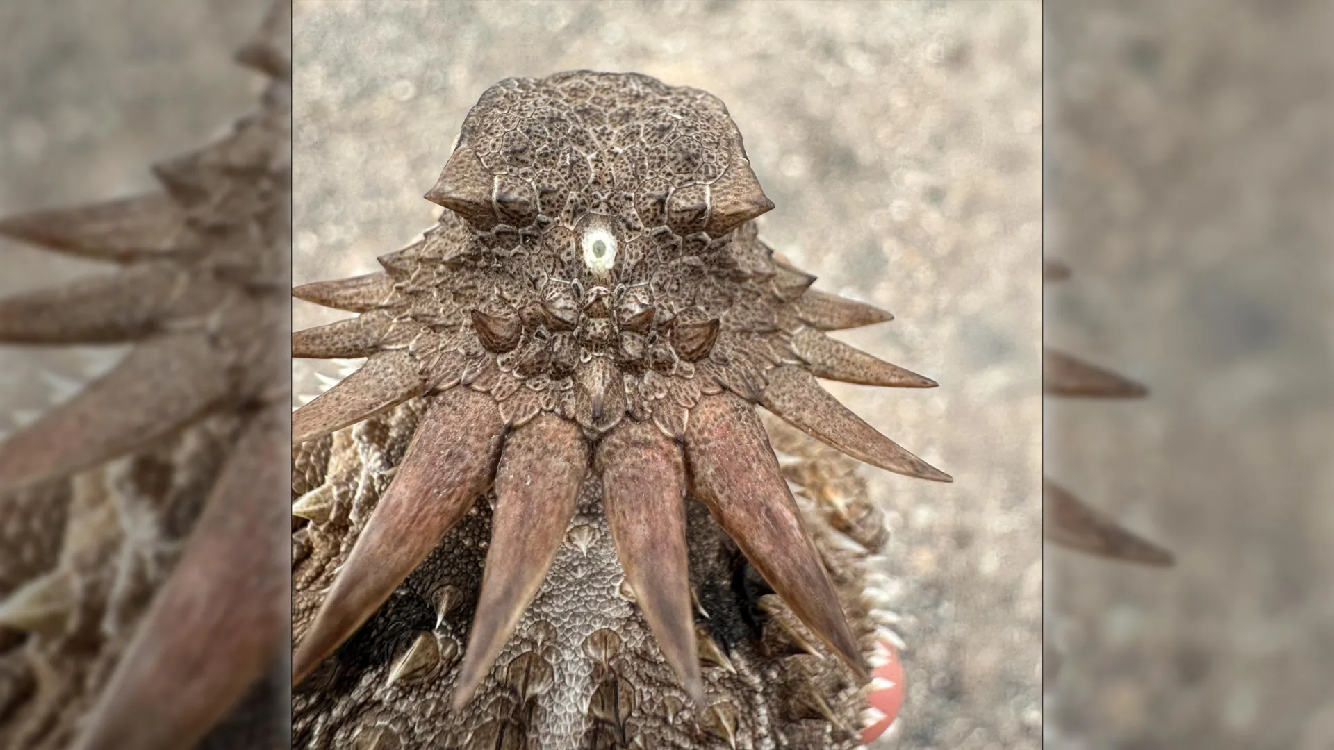
While human pineal glands do not form images, they retain direct biochemical links to light perception. In many lower vertebrates (such as certain lizards and amphibians), the pineal gland is literally a "third eye," covered only by a thin layer of skin and directly sensitive to sunlight. In mammals and humans, while light information is primarily captured by our paired retinal eyes, the signal is routed directly to the pineal gland to modulate the synthesis of melatonin—the master hormone governing our circadian rhythms.
When ambient light fades at eventide, retinal signals inform the brain, prompting the pineal gland to ramp up melatonin production, signaling to the body that it is time for rest. When dawn breaks, light suppression halts melatonin synthesis, waking up the organism. That our daily sleep cycle is governed by a structural descendant of a 600-million-year-old cyclopean sea creature is one of the most astonishing realizations in modern biology.
OFFICIAL STATEMENTS: PERSPECTIVES FROM THE RESEARCH FRONT
The findings, which challenge textbook dogmas regarding sensory evolution, have sent ripples through the international scientific community.
Prof. Dan-E Nilsson, professor emeritus in sensory biology at Lund University and a leading voice in the study of eye evolution, expressed profound astonishment at the conclusions drawn from the data:
"The results are a surprise. They turn our understanding of the evolution of the eye and the brain upside down," stated Nilsson during a press briefing discussing the research outcomes.
Highlighting the structural anomalies that have perplexed generations of anatomists, Nilsson emphasized how this model finally resolves a century-old biological debate:
"Now we finally understand why the eyes of vertebrates differ so radically from the eyes of all other animal groups, such as insects and squid. The film of our eyes—the retina—developed from the brain, whereas the eyes of insects and squid originate in the skin on the sides of the head."
Furthermore, Nilsson noted the breakthrough this provides regarding the neurological machinery of sight:
"For the first time, we now also understand the origin of the neural circuits that analyze the image in our retina."
Reflecting on the sheer improbability of our daily physiological experiences when viewed through an evolutionary lens, Nilsson concluded with a striking perspective:
"It’s mind-boggling that our pineal gland’s ability to regulate our sleep according to light stems from the cyclopean median eye of a distant ancestor 600 million years ago."
Collaborating researchers from the University of Sussex echoed these sentiments, pointing out that the methodology—combining molecular phylogenetics, embryology, and comparative histology—allows modern science to reconstruct soft-tissue anatomy that leaves virtually no direct fossil record. Because eyes and brains are soft tissues that rarely fossilize, piecing together their deep evolutionary history has traditionally required brilliant detective work. This new model provides a unified framework that bridges the gap between ancient genetic switches and modern physiological traits.
FUTURE OUTLOOK: WHERE RESEARCH GOES FROM HERE
As this study reshapes academic literature, it also opens up compelling new avenues for future scientific inquiry. The implications of tracing vertebrate eyes back to a repurposed median structure extend across multiple disciplines, from evolutionary developmental biology (evo-devo) to ophthalmology and neurology.
1. Mapping the Genetic Switches (Evo-Devo Insights)
One of the primary goals for subsequent research teams is to isolate the specific regulatory genes—such as the Pax6 master control gene for eye development—and track how their expression patterns shifted during the transition from a median eye to paired eyes. By utilizing advanced gene-editing tools like CRISPR-Cas9 in model organisms, researchers hope to experimentally replay minor loops of this evolutionary tape, observing how neural tissue reorganizes when embryonic pathways are nudged toward ancestral states.
2. Clinical Applications in Ophthalmology and Neurology
Understanding the deep embryological origins of the vertebrate retina provides fresh insights into congenital eye disorders and degenerative retinal diseases. Because the retina is fundamentally an extension of the central nervous system, conditions affecting retinal neurons often mirror neurodegenerative pathologies like Alzheimer’s and Parkinson’s disease. By mapping how neural circuits were originally assembled in our ancient ancestors, neuroscientists gain a clearer blueprint of how these complex networks function, potentially aiding the development of bioengineered retinal implants and neural regeneration therapies.
3. Broadening the Phyla Comparison
While the Lund-Sussex study bridges vertebrates and prominent invertebrates, researchers are expanding their comparative datasets to include obscure marine chordates, such as lancelets (amphioxus) and tunicates (sea squirts). These modern "living fossils" occupy crucial branching points near the root of the chordate family tree. Analyzing their light-detecting systems will help fine-tune the chronology of when the cyclopean stage occurred and whether similar visual detours happened in other unrecorded lineages.
Conclusion
The revelation that humans share a lineage with a tiny, one-eyed, filter-feeding creature from the Precambrian era is a humbling reminder of life’s winding, unpredictable path. Evolution does not operate as a neat, linear progression toward perfection; it is a master tinkerer, recycling, repurposing, and building upon the anatomical scraps of the past. The next time you gaze up at the stars, or feel the gentle pull of sleep as midnight approaches, remember that you are experiencing the legacy of a 600-million-year-old cyclops, gazing quietly out at an ancient world.