
Executive Overview
Long before the first fish swam, the first dinosaur walked, or human consciousness began pondering the cosmos, our deepest evolutionary ancestors may have stared at the ancient prehistoric oceans through a single, central eye.
In a paradigm-shifting study that upends decades of conventional zoological wisdom, researchers from Lund University and the University of Sussex have unveiled a startling conclusion: the earliest vertebrate ancestors passed through a "cyclops-like" stage nearly 600 million years ago. During this peculiar epoch of prehistoric life, these worm-like marine organisms relied entirely on a single median eye positioned centrally on top of their heads.
More remarkably, this new biological model suggests that this ancient, singular visual organ did not simply vanish into the evolutionary ether. Instead, it directly contributed to the genesis of the paired, image-forming eyes that humans and all other vertebrates use today. Even more astonishingly, a structural remnant of this primordial third eye continues to reside deep within the modern human brain as the pineal gland—the master regulator of our circadian rhythms and sleep cycles.
"The results are a surprise," says Dan-E Nilsson, professor emeritus in sensory biology at Lund University and a leading voice behind the research. "They turn our understanding of the evolution of the eye and the brain upside down."
This investigative feature explores the mechanics of this evolutionary detour, the comparative biology that separates vertebrates from invertebrates like insects and cephalopods, and the profound implications this discovery holds for our understanding of the human body.
Detailed Chronology: A 600-Million-Year Evolutionary Journey
To comprehend how humanity inherited its complex visual system, evolutionary biologists must look back to the Ediacaran and early Cambrian periods, an era when multicellular life was undergoing an explosive and chaotic diversification in Earth’s shallow seas.
Phase 1: The Loss of Paired Vision
According to the research team’s phylogenetic and comparative models, the distant predecessor to vertebrates was a modest, worm-like marine creature. Roughly 600 million years ago, this organism adopted a sedentary, benthic lifestyle. Rather than actively hunting or navigating complex terrain, it anchored itself to the seafloor, spending its days filtering plankton and organic detritus from the surrounding seawater.
Earlier in its lineage, this organism appears to have possessed paired light-sensitive cells or primitive proto-eyes. Throughout the animal kingdom, paired eyes are the evolutionary standard for mobile creatures, allowing brains to process binocular cues, calculate distance, judge direction, and map the positions of predators and prey.
However, evolutionary pressures are relentlessly pragmatic. When an animal abandons an active lifestyle in favor of a stationary, filter-feeding existence, maintaining complex neural and sensory architecture becomes metabolically expensive and functionally useless.
"We don’t know whether the paired eyes in our branch of the evolutionary tree were just light-sensitive cells or simple image-forming eyes," notes Professor Nilsson. "We only know that the organism later lost them."
Over countless generations, as the need for spatial awareness plummeted, the organism’s paired visual structures gradually degraded and disappeared from the genetic blueprint.
Phase 2: The Reign of the Median Eye
While the paired eyes faded away, total darkness was not an option for survival, even for a stationary filter-feeder. The organism retained a crucial cluster of photoreceptor cells positioned squarely in the middle of its head.
This cluster evolved into a functional median eye—a primitive, single-lens or pinhole-style visual organ. While incapable of resolving sharp images, detecting fine shapes, or tracking rapid motion, this central eye provided vital environmental cues:
- Circadian Awareness: It allowed the creature to distinguish the rhythm of day from night.
- Geotaxis/Orientation: It helped the animal determine which direction was upward toward the water’s surface, optimizing its feeding posture.
For millions of years, this cyclopean arrangement served the lineage well. The animal was adapted to its niche, content with a single eye to guide its static routines.
Phase 3: The Return to Mobility and the Rise of Vertebrate Eyes
As Earth’s ecological theater shifted, environmental pressures forced a behavioral revolution among these ancient organisms. Descendants of the sedentary filter-feeders abandoned their stationary habits and returned to an active, swimming lifestyle.
A mobile existence immediately resurrected an acute need for sophisticated vision. To navigate complex marine environments, hunt effectively, and evade fast-swimming predators, these animals required advanced image-forming eyes once more.
Rather than reinventing vision from scratch, evolution took an ingenious, albeit bizarre, detour. The researchers conclude that portions of the original median eye were dynamically repurposed and expanded. Through unprecedented genetic and morphological remodeling, this central visual structure spawned a brand-new pair of lateral eyes capable of sophisticated image formation.
This roundabout evolutionary history solves a long-standing biological mystery: why vertebrate eyes are constructed so fundamentally differently from the eyes of other successful animal groups, such as insects, spiders, and cephalopods like squid and octopuses.
"Now we finally understand why the eyes of vertebrates differ so radically from the eyes of all other animal groups," explains Nilsson. "The film of our eyes—the retina—developed from the brain, whereas the eyes of insects and squid originate in the skin on the sides of the head."
Supporting Context & Metrics: Decoding the Anatomical Divide
The divergence in eye origin between vertebrates and invertebrates represents one of the most profound structural partitions in evolutionary biology. To understand the weight of the Lund-Sussex findings, one must examine the micro-anatomy and embryology of vision.
The Embryonic Origin of the Retina
In human beings and all other vertebrates, the retina—the delicate, neural tissue lining the interior back of the eyeball—is an direct extension of the central nervous system. During embryonic development, retinal tissue bulges outward from the developing brain, bringing complex neural processing power directly to the optical frontline.
Conversely, invertebrate eyes—such as the compound eyes of a housefly or the camera-like eyes of a giant squid—arise from the ectoderm, the outer embryonic layer that forms the skin. In these creatures, light-detecting cells fold inward from the surface of the head, connecting backward to the brain via peripheral nerves later in development.
| Feature | Vertebrate Eyes (Humans, Fish, Birds) | Invertebrate Eyes (Insects, Cephalopods) |
|---|---|---|
| Embryonic Origin | Outgrowth of brain tissue (neuroectoderm) | Outgrowth of surface skin tissue (surface ectoderm) |
| Retinal Development | Forms internally as part of the central nervous system | Forms externally via invagination of the integument |
| Neural Circuitry | Pre-processes visual signals within the retina before transmission | Relies heavily on post-retinal brain centers for early parsing |
| Evolutionary Lineage | Derived via repurposing of an ancient median eye | Evolved independently via surface photoreceptor clusters |
The Mechanics of Retinal Neural Circuits
This unique evolutionary pathway also explains the origin of the sophisticated neural circuits embedded within the vertebrate retina. Because the retina is essentially brain tissue pushed outward, it does not merely act as a passive camera film capturing light photons.
Instead, specialized retinal interneurons (such as bipolar, amacrine, and ganglion cells) begin analyzing visual data before the electrical signals even reach the visual cortex of the brain. These localized networks parse brightness gradients, edge contrasts, chromatic information, and directional motion natively.
"For the first time, we now also understand the origin of the neural circuits that analyze the image in our retina," Professor Nilsson observes. These circuits are the direct evolutionary inheritance of a central processing node that once managed simple light cues for our one-eyed ancestors.
Official Statements & Scientific Methodology
The conclusions put forth by the Lund University and University of Sussex research teams are not speculative assumptions; they are built upon an exhaustive comparative framework.
To trace the lineage of light-detecting cells, the international research team mapped the distribution, genetic markers, functional properties, and neural connectivity of photoreceptors across a vast cross-section of modern and extinct animal phylogenies. By comparing how these cells wire into surrounding tissues, the team reconstructed the most probable sequence of evolutionary transitions.
"The evidence suggests a very specific, step-by-step sequence of structural changes," notes a co-researcher associated with the study. "We moved from dual primitive spots to a streamlined, single median eye due to disuse, only to experience an unprecedented evolutionary exaptation—recycling parts of that median architecture to build the advanced lateral eyes we see today."
The Living Fossil Within: The Pineal Gland
Perhaps the most captivating dimension of this research involves the fate of the median eye itself. While the main optical components were repurposed to build paired lateral eyes, the original median eye did not completely vanish.
Instead, deep within the center of the modern vertebrate brain sits a small, pinecone-shaped endocrine organ: the pineal gland.
While humans and mammals do not use the pineal gland to perceive visual images of the external world, its deep physiological roots remain tethered to ancient light-sensing duties. The pineal gland is chiefly responsible for synthesizing and secreting melatonin, a critical hormone that regulates circadian rhythms—the internal biological clocks that dictate sleep-wake cycles across a 24-hour solar day.
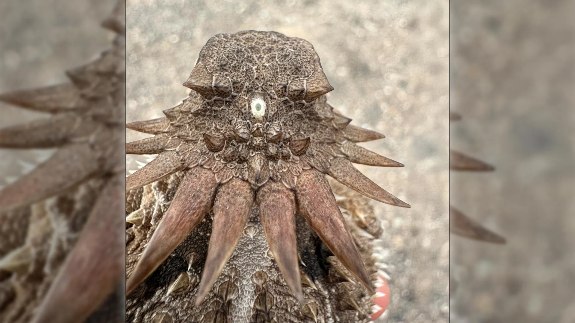
In many lower vertebrates, such as certain fish, amphibians, and reptiles, the pineal complex (sometimes referred to as the "parietal eye" or "third eye") remains directly sensitive to ambient sunlight, sitting just beneath a thin, translucent patch of skull bone.
In humans, while the pineal gland is no longer directly exposed to light, it receives photic signals indirectly via complex neural pathways originating in the retinal cells of our paired eyes. As evening falls and light levels drop, retinal signals trigger the pineal gland to ramp up melatonin production, inducing drowsiness. As dawn breaks, light suppression halts melatonin synthesis, promoting alertness.
"It’s mind-boggling that our pineal gland’s ability to regulate our sleep according to light stems from the cyclopean median eye of a distant ancestor 600 million years ago," Professor Nilsson concludes.
Future Outlook & Implications for Evolutionary Biology
The revelation that vertebrates passed through a one-eyed, cyclopean phase opens exciting new avenues for paleobiology, developmental genetics, and neurobiology. As researchers continue to map the genetic switches that control embryonic tissue differentiation, testing the molecular pathways of median-eye development could yield profound insights into congenital eye disorders and evolutionary developmental biology (evo-devo).
Furthermore, this study serves as a stark reminder of the wild, non-linear trajectories of evolution. Popular conceptions of evolution often imply a steady, continuous march from simple to complex. Yet this research demonstrates that evolution is entirely opportunistic—capable of discarding complex organs when lifestyles change, and equally capable of salvaging, repurposing, and re-engineering ancient structures to meet new environmental demands.
The next time you gaze up at the night sky, or feel the heavy pull of sleep as midnight approaches, consider the journey of your cellular lineage. Deep within your skull, a tiny gland quietly processes the passage of light and dark, operating via a biological script written in the Ediacaran seas—a lingering echo from a time when your earliest ancestor faced the vast ocean with a single, unblinking eye.