
Executive Overview
As winter descends, bringing with it the familiar and unwelcome triad of fever, aching limbs, and a runny nose, public health systems brace annually for the onslaught of seasonal influenza. Caused by highly adaptable influenza viruses that invade the human body primarily through airborne respiratory droplets, the disease remains a formidable global health challenge. Millions of cases are recorded each year, leading to hundreds of thousands of hospitalizations and placing immense strain on healthcare infrastructure worldwide. Despite decades of study, the precise, dynamic mechanics by which the influenza virus bridges the extracellular environment and the interior of a living human cell have long remained obscured by the limitations of traditional scientific instrumentation.
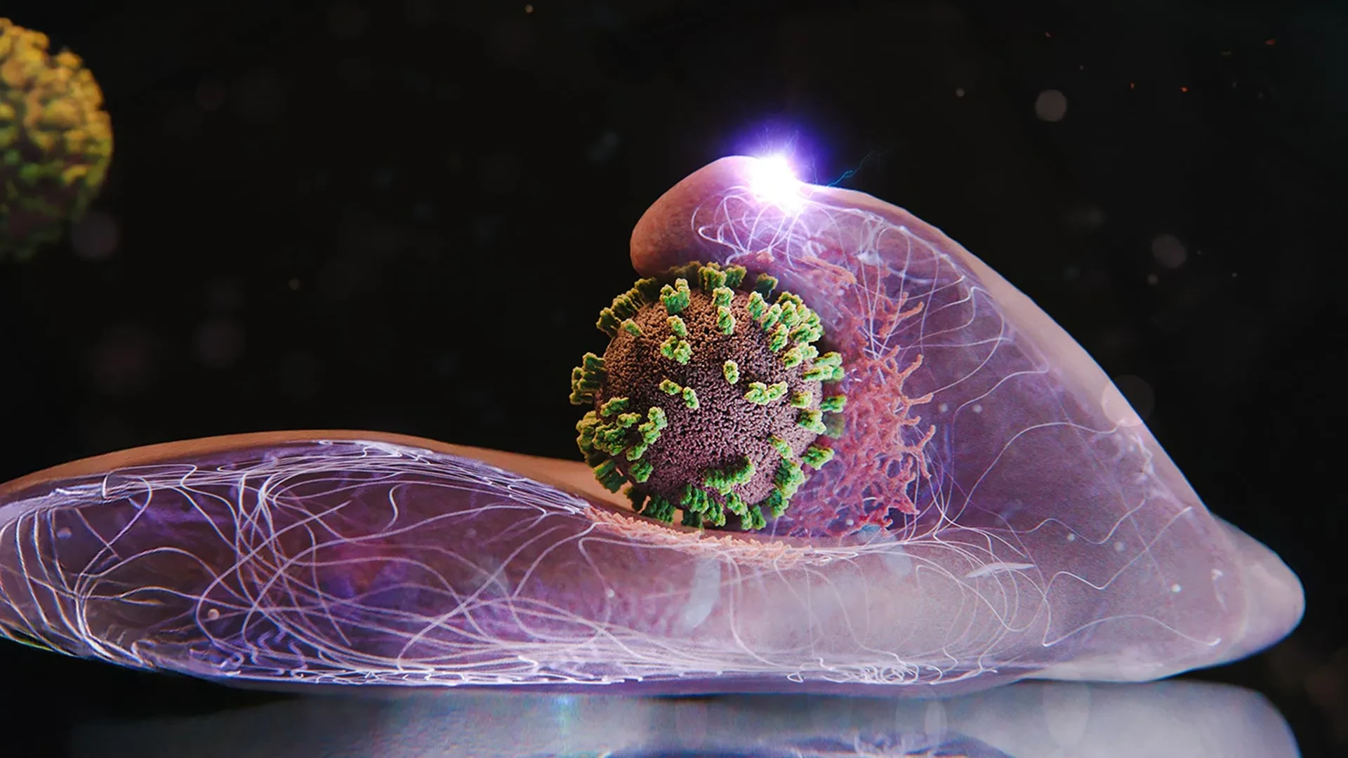
Now, a pioneering collaborative research team hailing from Switzerland and Japan has pierced this microscopic veil. Under the visionary direction of Yohei Yamauchi, Professor of Molecular Medicine at ETH Zurich, the consortium has developed an unprecedented microscopy technique that allows scientists to zoom in on the outer surface of living human cells cultivated in a Petri dish. By combining advanced visualization modalities, the researchers have successfully watched—live, in real time, and with breathtaking spatial detail—the exact moment an influenza virus penetrates a host cell.
The implications of this breakthrough stretch far beyond academic curiosity. Far from being a passive victim in this microscopic drama, the host cell actively participates in its own subversion. Observing this process has led Professor Yamauchi to describe the infection not as a simple breach, but rather as an intricate, coordinated "dance between virus and cell."
This article explores the mechanics of this viral infiltration, the cutting-edge technology that made its observation possible, the deeper biological insights uncovered by the ETH Zurich team, and how these findings pave the way for a revolutionary generation of antiviral therapeutics and vaccines.
Detailed Chronology: The Step-by-Step Mechanics of Viral Entry
To comprehend the magnitude of the ETH Zurich and Japanese research team’s findings, one must first trace the chronological sequence of events that unfolds when an influenza virus encounters a susceptible human cell. Using their newly minted visualization platform, the scientists were able to break down the infection process into distinct, observable phases.
Phase 1: Attachment and Membrane Surfing
The initial encounter between the influenza virus and a human cell is governed by molecular affinity. The virus particle, or virion, carries surface glycoproteins—specifically hemagglutinin—that seek out and bind to specific sialic acid receptors embedded in the plasma membrane of the host cell.
However, the virus does not merely land and anchor. Instead, the research team observed a phenomenon they have termed "viral surfing." Once attached, the virus moves dynamically across the cell surface. It engages in a continuous process of unbinding and rebinding, latching onto one receptor molecule after another. This surface migration is not random; the virus effectively navigates along a biochemical gradient, driven toward localized membrane zones that are densely populated with the necessary receptors. By migrating to a receptor-rich hotspot, the virus maximizes its chances of successful internalization.
Phase 2: Cellular Co-optation and Active Sequestration
Once the virus reaches an optimal entry site, the dynamic relationship shifts from passive surfing to active cellular engagement. Conventional wisdom in virology once held that viruses simply trick cells into engulfing them by mimicking benign extracellular cargo. However, the live-imaging data captured by Yamauchi’s team revealed a surprising twist: the host cells do not sit idle.
Instead, the cells make an active effort to seize the incoming pathogen. The interaction looks deceptively cooperative because the virus exploits a routine, indispensable cellular uptake system known as receptor-mediated endocytosis. This internal transport mechanism is vital for normal cellular physiology, responsible for ferrying essential metabolic substances—such as hormones, cholesterol, and iron—into the cell’s interior.
As the virus lingers at a receptor-dense zone, the host cell’s plasma membrane responds. The membrane begins forming a localized indentation directly beneath the viral particle. Simultaneously, the cell summons specialized structural proteins, chief among them clathrin, to the immediate site of attachment. Clathrin molecules polymerize to form a scaffolding network that shapes and physically supports the deepening pocket in the membrane.
Phase 3: Encasement and Internalization
As the clathrin-coated pit deepens, it progressively wraps around the influenza virus, eventually pinching off from the inner surface of the plasma membrane to form a closed intracellular container known as a vesicle (or endosome).
The cell then actively pulls this vesicle inward, drawing the trapped virus deep into the cytoplasm. Once safely inside the intracellular environment, the protective clathrin coat disassembles and dissolves. This uncoating process exposes the viral core to the changing biochemical conditions of the endosome, ultimately allowing the genetic material of the influenza virus to escape into the host cell machinery, where it commands the cell to mass-produce copies of the pathogen.
Crucially, the ETH Zurich researchers noted that this process is characterized by active defensive or exploratory maneuvers by the cell. When the virus attempts to drift away from the membrane during the initial attachment phase, the cell’s surface exhibits distinct, wave-like motions, pushing upward as if actively trying to corral and capture the elusive particle.
Supporting Context & Metrics: Overcoming Historical Limitations
The breakthrough achieved by Professor Yamauchi and his colleagues was not merely a matter of observation, but of technological innovation. For decades, virologists labored under severe methodological constraints that restricted their understanding of viral entry to static snapshots or low-resolution approximations.
Why Earlier Microscopy Fell Short
To appreciate the significance of the new imaging platform, one must examine the shortcomings of legacy techniques:
- Transmission Electron Microscopy (TEM): Historically, electron microscopy provided the highest spatial resolution available to researchers, enabling detailed structural analyses of viruses and cellular membranes. However, TEM requires rigorous sample preparation, including chemical fixation, dehydration, and heavy-metal staining. These procedures inevitably kill the biological specimen. Consequently, electron microscopy can only capture static, dead moments in time—akin to taking a single photograph of a dynamic car crash and attempting to reconstruct the physics of the entire collision.
- Traditional Fluorescence Microscopy: While fluorescence microscopy permits the observation of live cellular processes in real time, it suffers from a major drawback: low spatial resolution. The diffraction limit of light makes it difficult to resolve fine-scale membrane dynamics and the precise nanometer-level interactions occurring between a single virion and the cell surface.
The Birth of ViViD-AFM
To bridge the gap between high spatial resolution and real-time live imaging, the research team engineered a hybrid methodology. They successfully merged Atomic Force Microscopy (AFM) with advanced fluorescence microscopy, creating an integrated apparatus christened virus-view dual confocal and AFM (ViViD-AFM).
Atomic force microscopy operates on an entirely different physical principle than light- or electron-based optics. An AFM system uses an extraordinarily sharp microscopic probe—a nanoscale tip mounted on a flexible cantilever—to physically scan across the surface of a sample. By measuring the minute deflections of the cantilever as the tip interacts with the surface forces of the living cell, the instrument can generate a three-dimensional topographical map with nanometer-scale precision.
By synchronizing this topographical scanning with high-sensitivity confocal fluorescence microscopy, the ViViD-AFM platform allows scientists to visualize biochemical markers (via fluorescence) while simultaneously tracking the exact physical contours and mechanical deformations of the cell membrane (via AFM) in real time. This dual-modal approach provided the ETH Zurich and Japanese team with the unprecedented clarity needed to watch the cellular ballet of viral entry unfold before their eyes.
Official Statements and Expert Insights
The implications of the ViViD-AFM technology and the unexpected discoveries regarding cell-virus interactions have generated significant commentary within the scientific community.
Reflecting on the nature of the discovery, lead investigator Professor Yohei Yamauchi emphasized the paradigm shift in how immunologists and virologists must view cellular susceptibility.
"The infection of our body cells is like a dance between virus and cell," stated Professor Yamauchi, Molecular Medicine expert at ETH Zurich.
This metaphor underscores the active, reciprocal nature of the infection process. Rather than acting as a passive wall breached by an aggressive invader, the human cell is an unwitting participant, drawn into a choreographed sequence of biochemical handshakes and mechanical responses.
Furthermore, members of the research consortium have highlighted the collaborative nature of the breakthrough, noting that bridging the disciplines of biophysics, molecular biology, and advanced optical engineering was essential. By uniting Swiss and Japanese expertise, the team was able to overcome technical hurdles that had stymied individual laboratories for years.
Independent virologists not affiliated with the study have praised the ViViD-AFM technique as a methodological leap forward. By moving beyond the binary choice between high-resolution static imaging and low-resolution dynamic imaging, the scientific community now possesses a versatile lens through which to examine cellular pathology at the nanoscale.
Future Outlook: Implications for Antiviral Research and Global Health
The development of the ViViD-AFM platform and the granular insights gained into influenza entry open up vast new horizons for biomedical research, drug discovery, and preventive medicine.
Direct Evaluation of Antiviral Drug Candidates
Traditionally, the screening of antiviral drugs has relied heavily on downstream assays—measuring whether a viral population survived or perished hours or days after drug administration. While effective, these assays reveal little about how a drug interfered with the infection cycle at the physical level.
Because ViViD-AFM permits the real-time observation of infection as it happens in live cell cultures, it provides pharmaceutical researchers with a direct, high-resolution assay platform. Scientists can now introduce candidate antiviral compounds into the Petri dish and watch, minute by minute, whether the drug successfully blocks viral surface surfing, prevents clathrin recruitment, or halts membrane indentation. This capability allows for the rapid identification and optimization of entry inhibitors—drugs designed to stop the virus before it ever crosses the cellular threshold.
Applications Beyond Influenza
While the initial studies focused on the influenza virus, the foundational mechanics of viral entry—such as receptor recruitment, membrane surfing, and clathrin-mediated endocytosis—are utilized by a wide array of human pathogens. The research team notes that the ViViD-AFM technique is highly adaptable. It can readily be deployed to study the cellular entry mechanisms of other dangerous viruses, ranging from respiratory syncytial virus (RSV) to coronaviruses and beyond.
Moreover, the technology holds significant promise for vaccine research. By offering a real-time view of how viral particles, virus-like particles (VLPs), or novel vaccine delivery vectors interact with human cells, immunologists can refine the design of vaccines to ensure optimal cellular uptake and immune stimulation.
Conclusion
As seasonal influenza continues to pose a persistent threat to global public health, the work being conducted at ETH Zurich and partner institutions in Japan offers renewed hope. By illuminating the intricate, dynamic choreography between invading pathogens and human cells, researchers are transforming our understanding of infectious disease. Armed with tools like ViViD-AFM, the scientific community is better equipped than ever to outmaneuver the flu, turning the tables on a microscopic dance that has challenged humanity for generations.