
Executive Overview
For decades, the evolutionary journey of vertebrate vision was taught as a relatively straightforward tale of gradual refinement. Biologists understood that our ancient aquatic forebears developed paired ocular organs to navigate the primordial seas, steadily sharpening their focus through natural selection until these structures evolved into the complex cameras we possess today. However, groundbreaking new research spearheaded by collaborative teams at Lund University and the University of Sussex has shattered this conventional paradigm.
According to their findings, the lineage of every living vertebrate—from deep-sea fish to birds, reptiles, and human beings—traces back to a bizarre, worm-like organism that lived nearly 600 million years ago. This distant relative was, in effect, a biological cyclops. It abandoned its original paired eyes in favor of a single, median optical organ positioned squarely on top of its head.
This startling revelation does more than merely add a peculiar footnote to evolutionary biology; it completely upends our understanding of how both the vertebrate eye and brain developed. Most notably, the research reveals that this ancient, single-eyed design did not vanish into the annals of prehistory. Its literal remains persist inside the human skull to this day: the pineal gland, a tiny, pinecone-shaped endocrine structure buried deep within the brain that regulates our sleep-wake cycles.
By analyzing the deep genetic and structural architectures of light-sensitive cells across diverse animal groups, scientists have solved a long-standing evolutionary mystery. They have finally answered why vertebrate eyes are fundamentally constructed in a manner entirely distinct from those of invertebrates like insects and cephalopods, while simultaneously uncovering the bizarre, roundabout path that gave rise to human consciousness and vision.
Detailed Chronology of an Evolutionary Detour
To comprehend how modern human vision originated from a microscopic, cyclopean worm, one must journey backward through deep time to the Ediacaran period, roughly 600 million years ago.
Phase I: The Stationary Settler and the Loss of Sight
During this epoch, the Earth’s oceans hosted a burgeoning array of soft-bodied, primitive multicellular organisms. Among them was a modest, worm-like creature that anchored itself to the seafloor or sediment, feeding passively by filtering microscopic plankton from the surrounding seawater.
According to evolutionary biologist Professor Emeritus Dan-E Nilsson of Lund University, this creature’s earliest ancestors likely possessed two paired eyes—similar to many other contemporary aquatic species. However, as the organism adapted to an increasingly sedentary, stationary lifestyle, the evolutionary utility of maintaining complex, paired eyes plummeted.
In the calculus of natural selection, maintaining unused sensory organs metabolic costs energy. Consequently, over countless generations, this branch of the evolutionary tree discarded its paired eyes. The organism became functionally blind in the traditional sense, relying instead on simpler tactile and chemical cues to survive its immobile existence.
Phase II: The Rise of the Median Eye
Yet, nature rarely abandons light-sensing capabilities entirely. Even after the paired eyes vanished, a small cluster of primitive, light-sensitive cells remained preserved in the exact center of the creature’s head.
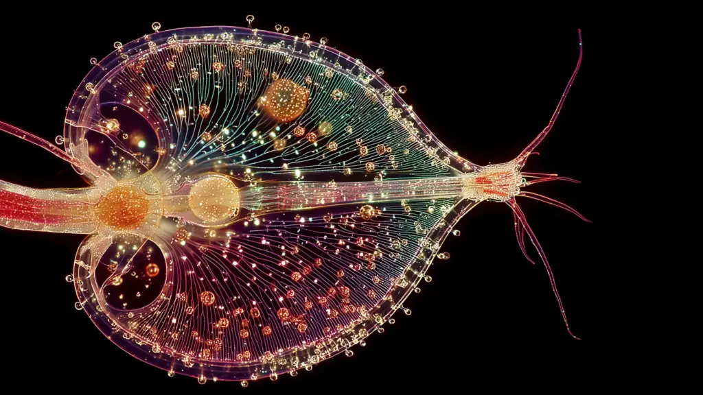
Over millions of years, these retained cells coalesced and evolved into a simple, single "median eye." While this rudimentary structure was incapable of forming crisp images, it served a vital survival function: it allowed the organism to detect the broad shifts between day and night, sense looming shadows from predators overhead, and maintain a basic sense of spatial orientation relative to the sun. This tiny, centralized dot of photoreceptors made the creature a literal aquatic cyclops.
Phase III: The Return to the Swim and the Rebuilding of Vision
The evolutionary plot thickened dramatically when ecological pressures shifted once again. Millions of years after adopting a stationary filter-feeding existence, descendants of this worm-like ancestor abandoned their sedentary habits and returned to a fully active, swimming lifestyle.
This profound behavioral pivot triggered an immediate, intense evolutionary demand for advanced sensory perception. Navigating open waters, avoiding agile predators, and hunting dynamic prey required sophisticated vision.
Rather than reinventing ocular organs from scratch or somehow resurrecting the long-lost paired eyes of its more distant ancestors, evolution took an astonishing detour. The organism repurposed and expanded upon the existing median eye. Parts of this single central structure gradually proliferated and migrated, eventually giving rise to the new, paired, image-forming eyes that became the hallmark of the vertebrate subphylum.
Supporting Context & Metrics: The Architectural Divide in Animal Vision
To appreciate the weight of this discovery, one must examine the profound structural gulf that separates vertebrate eyes from those of invertebrates. For generations, comparative anatomists have marveled at a fundamental embryological contradiction:
- Vertebrate Eyes (Humans, Birds, Fish): The retina—the delicate, light-sensitive film lining the back of the eye—develops directly out of embryonic brain tissue. In a very literal sense, human eyes are protruding extensions of the central nervous system.
- Invertebrate Eyes (Insects, Spiders, Squid, Octopus): The light-detecting tissues develop entirely from the ectoderm, the outer embryonic layer that forms the animal’s skin on the sides of the head.
For decades, textbook explanations struggled to reconcile how two entirely different lineages could arrive at sophisticated camera-type eyes using such radically divergent developmental pathways.
The new findings from Lund University and the University of Sussex provide the missing puzzle piece. Because vertebrate vision was rebuilt from a central median eye that sat atop the head and was intimately tied to the neural architecture of the nascent brain, its developmental blueprint remained tethered to the central nervous system. In contrast, invertebrates like insects and squid followed separate evolutionary lineages that developed epidermal photoreceptors on the flanks of their bodies.
Furthermore, this discovery sheds light on the deep evolutionary origins of neural circuits. For the first time, researchers can trace how the complex neural machinery responsible for processing visual data within the human retina originally assembled around that ancient, single-eyed organ 600 million years ago.
Official Statements and Academic Insights
The implications of this research are sending ripples through the global scientific community. In joint statements released following the publication of their findings, the research team emphasized just how radical a shift this represents for evolutionary theory.
"The results are a surprise. They turn our understanding of the evolution of the eye and the brain upside down," stated Dan-E Nilsson, professor emeritus in sensory biology at Lund University.
Nilsson, whose career has focused extensively on the comparative mechanics of sensory organs, noted that piecing together this evolutionary puzzle required looking beyond gross anatomy and diving deep into cellular and genetic comparisons across diverse animal phyla.
"We don’t know whether the paired eyes in our branch of the evolutionary tree were just light-sensitive cells or simple image-forming eyes," Nilsson explained, addressing the transitional gaps in the Ediacaran fossil record. "We only know that the organism later lost them… For the first time, we now also understand the origin of the neural circuits that analyze the image in our retina."
Co-researchers from the University of Sussex echoed these sentiments, highlighting the sheer improbability of the evolutionary path taken. The transition from an active ancestor with two eyes to a stationary, one-eyed filter feeder, followed by a return to active swimming that required rebuilding vision from a median photoreceptor, demonstrates that evolution is not a straight ladder of progress, but a labyrinth of opportunistic recycling.
The Pineal Gland: A Living Fossil in the Human Brain
Perhaps the most captivating aspect of this discovery for the general public is the direct bridge it builds between deep evolutionary history and modern human anatomy.
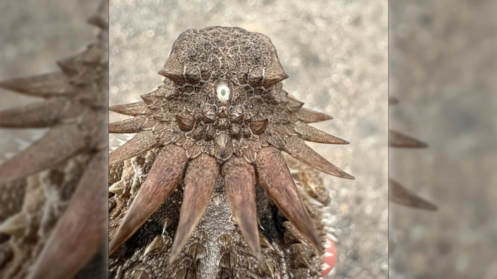
The single median eye of that ancient, worm-like creature did not completely disappear when vertebrates evolved paired, image-forming eyes. Instead, it was sequestered deep within the cranium, evolving into what we now call the pineal gland.
Located near the center of the brain, nestled between the two hemispheres, the pineal gland is a tiny, pinecone-shaped endocrine organ. While it is no longer exposed to direct sunlight in mammals, it retains a deep, ancient biochemical connection to light.
In humans and other vertebrates, the pineal gland functions as the body’s internal pacemaker. It synthesizes and secretes melatonin, a hormone regulated directly by the photoperiod—the cycle of light and darkness in our environment. When darkness falls, our eyes signal the brain, prompting the pineal gland to release melatonin, which induces drowsiness and orchestrates our circadian rhythms.
"It’s mind-boggling that our pineal gland’s ability to regulate our sleep according to light stems from the cyclopean median eye of a distant ancestor 600 million years ago," reflects Nilsson.
This revelation transforms the pineal gland from a mysterious endocrine gland into a living fossil—a microscopic monument in our heads testifying to a time, hundreds of millions of years ago, when our earliest ancestors navigated the primordial seas guided by a single eye on the tops of their heads.
Future Outlook and Ongoing Research
As evolutionary biologists and neuroscientists digest these findings, the research is opening up vibrant new avenues of inquiry.
1. Genomic Mapping of Ancient Photoreceptors
Future studies will likely focus on mapping the precise molecular and genetic signatures of light-sensitive cells in primitive living chordates, such as lancelets and tunicates. By comparing the gene expression networks active in the pineal gland with those found in the retinas of vertebrates and the simple photoreceptors of invertebrates, researchers hope to reconstruct the exact genetic toolkit that enabled this evolutionary sleight-of-hand.
2. Paleontological Re-examination
Paleontologists are now taking a closer look at Ediacaran and early Cambrian microfossils. Armed with the hypothesis that early chordates underwent a cyclopean phase, researchers are re-evaluating poorly understood fossilized impressions for signs of median cephalic structures that were previously dismissed as preservation artifacts or unrelated anatomical features.
3. Broadening Medical and Chronobiological Understanding
Beyond pure evolutionary theory, a deeper understanding of the pineal gland’s ancestry may offer fresh perspectives in chronobiology. By tracking how light-sensing and neuroendocrine pathways have been conserved or modified across 600 million years, medical researchers can gain novel insights into circadian disorders, seasonal affective disorder (SAD), and the complex hormonal cascades that govern human sleep architecture.
Ultimately, this study serves as a humbling reminder of nature’s boundless ingenuity. The eyes through which we read these words, and the internal clock that tells us when to sleep, are the direct descendants of a blind-alley detour taken by a tiny, one-eyed worm in the twilight of the Precambrian world.