
Executive Overview
For decades, the standard textbook model of vertebrate evolution has maintained a relatively straightforward trajectory: as ancestral life crawled, swam, and adapted across shifting prehistoric landscapes, visual systems scaled in complexity from simple light-sensitive patches to advanced, dual image-forming organs. However, groundbreaking new research spearheaded by evolutionary biologists at Lund University and the University of Sussex has upended this neat narrative.
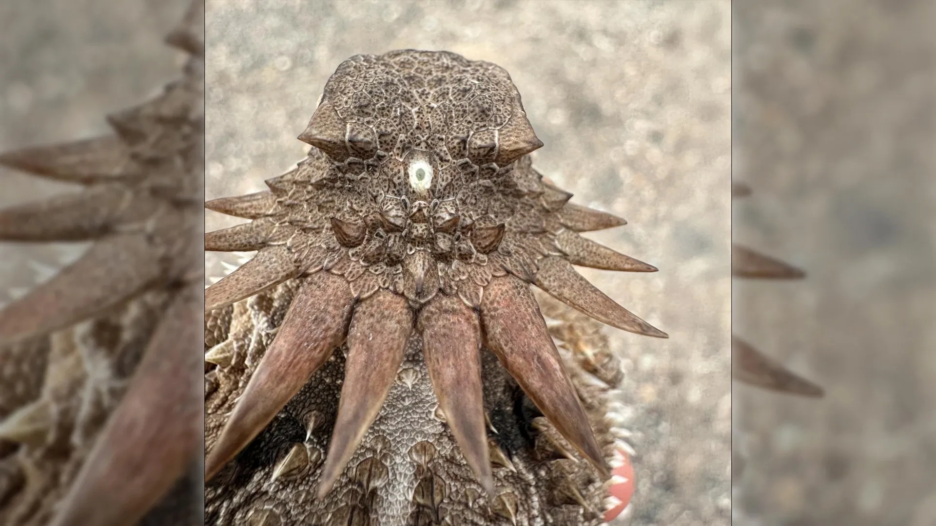
According to their findings, humanity—alongside every other living vertebrate, from the sparrow in the sky to the blue whale in the abyss—shares a surprisingly bizarre common ancestor: a tiny, worm-like creature that lived roughly 600 million years ago and possessed a single, central eye positioned atop its head, much like a microscopic cyclops.
This startling revelation does more than just inject a touch of mythological wonder into paleontology; it fundamentally re-evaluates the architecture of the human brain. The research reveals that the ancient "median eye" of our prehistoric forebear did not simply vanish into the mists of deep time. Instead, its cellular remnants persist today, hidden deep within the human cranium as the pineal gland—the neuroendocrine organ responsible for regulating our sleep-wake cycles and circadian rhythms. Furthermore, this bizarre evolutionary detour explains an enduring biological mystery: why vertebrate eyes are constructed so radically differently from those of invertebrates like insects and squids, with our retinas developing directly out of brain tissue rather than surface skin.
Detailed Chronology: The Twist and Turns of Deep-Time Vision
To understand how a single-eyed, sessile organism gave rise to the complex visual apparatus of modern vertebrates, researchers had to reconstruct a timeline spanning hundreds of millions of years, moving from sedentary filter-feeders to active swimmers.
Phase 1: The Pre-Cyclopean Baseline (Pre-600 Million Years Ago)
Long before the dawn of backboned animals, the earliest branches of the deuterostome lineage likely experimented with vision in various forms. While the exact ancestral state remains veiled by the incompleteness of the fossil record, scientists believe these primitive aquatic organisms possessed rudimentary, paired light-sensitive cells or simple eyespots, much like many contemporary marine invertebrates. These early tools were adequate for detecting the broad sweep of day and night, offering primitive survival advantages against ancient predators.
Phase 2: The Stationary Bottleneck and the Loss of Paired Eyes
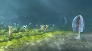
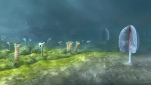
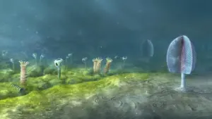
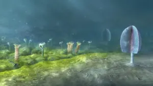
Approximately 600 million years ago, during the Ediacaran or early Cambrian periods, a specific branch of our distant ancestors adopted a drastically different ecological strategy. Rather than actively hunting or navigating complex terrain, this worm-like organism transitioned to a largely stationary, sedentary lifestyle. Anchoring itself to the substrate, it survived primarily by filtering plankton and organic particulate matter out of passing seawater.
In the evolutionary calculus of nature, maintaining complex physiological structures that offer no active survival benefit is an energetic waste. Because a stationary, filter-feeding organism did not need to hunt prey or evade dynamic threats using spatial vision, the evolutionary pressure to maintain paired lateral eyes vanished. Over generations, those paired structures atrophied and disappeared entirely.
Phase 3: The Rise of the Median "Cyclops" Eye
Yet, light detection remained useful even for a stationary filter-feeder—if only to track seasonal cycles, day-night shifts, or passing shadows. Crucially, even after the loss of the paired lateral eyes, a localized cluster of light-sensitive cells persisted in the exact center of the organism’s head.
Over immense spans of time, natural selection honed this cluster into a functional, singular median eye. Unlike image-forming eyes, this structure functioned primarily as a light-and-dark detector, helping the organism orient itself within its environment and synchronize its internal metabolic processes with ambient environmental cues. For a significant epoch in vertebrate prehistory, our lineage was, effectively, a population of tiny, aquatic cyclopes.
Phase 4: The Return to the Swimsphere and the Rebirth of Vision
Evolution is rarely a straight line, and stasis is seldom permanent. Millions of years after adopting a sedentary filter-feeding existence, ecological pressures shifted once again. Descendants of this worm-like organism broke free from their anchors, returning to a dynamic, active, swimming lifestyle in the primordial oceans.
This return to mobility instantly reinstated the evolutionary pressure for advanced vision. Navigating a complex three-dimensional watery world, hunting moving prey, and evading sophisticated predators required far more than a simple light-dark sensor. Intriguingly, rather than re-evolving eyes from scratch or resurrecting the long-lost lateral organs, evolution improvised. Parts of the ancient median eye and its associated neural pathways expanded, migrated, and re-differentiated, ultimately giving rise to the new, paired, image-forming eyes that define modern vertebrates.
Supporting Context & Metrics: Anatomy, Embryology, and the Invertebrate Divide
The implications of this discovery extend far beyond paleontological curiosity; they solve a foundational puzzle in comparative embryology that has vexed biologists for over a century: the vertebrate-invertebrate eye dichotomy.
The Embryological Divide: Brain vs. Skin
If one compares the eye of a human to the eye of a housefly or a colossal squid, the functional similarities can be deceptive. Both systems focus light through a lens onto a light-sensitive sheet of tissue to form crisp visual representations of the world. However, their developmental origins are entirely foreign to one another.
- Invertebrates (Insects, Mollusks, and Cephalopods): Their eyes develop from the ectoderm—specifically, from the surface skin cells on the sides of the head. The embryonic tissue folds inward or forms vesicles that differentiate into the retina and associated structures.
- Vertebrates (Fish, Amphibians, Reptiles, Birds, and Mammals): Our eyes originate directly from the central nervous system. The vertebrate retina is, quite literally, an outward extension of the embryonic brain that loops forward during development.
For generations, this structural gulf suggested that eyes had evolved completely independently multiple times across the animal kingdom. However, the new Lund-Sussex findings reconcile this paradox. Because vertebrate vision was rebuilt from a central median eye that was already intimately connected to the neural architecture of the head, the resulting visual organs naturally developed as extensions of the brain. The retina’s intricate neural circuits—the processing power that breaks down shapes, colors, and motion—were inherited directly from the ancient neural machinery that once serviced the cyclopean median eye.
The Pineal Gland: A Living Fossil in the Human Brain
Perhaps the most poetic and tangible proof of this evolutionary history sits quietly at the geometric center of the modern human brain: the pineal gland.
Shaped roughly like a tiny pinecone (hence its name), this endocrine gland has long been recognized for its crucial role in human physiology. Operating largely in the dark, the pineal gland synthesizes and secretes melatonin—a hormone driven by the suprachiasmatic nucleus of the hypothalamus that translates environmental light cues (or the lack thereof) into biochemical signals governing sleep cycles, seasonal behaviors, and circadian rhythms.
Under the lens of the new phylogenetic models, the pineal gland is cast in an entirely new light. It is not merely a specialized endocrine organ; it is a direct, living evolutionary remnant of the ancient median eye. Though it lost its lens and its ability to form images millions of years ago, the pineal gland retains its ancestral sensitivity to light-dark cycles, serving as an unbroken biochemical bridge connecting modern human consciousness to the watery world of our 600-million-year-old single-eyed ancestors.
Official Statements and Expert Analysis
The research, which bridges sensory biology, genomics, and evolutionary developmental biology (evo-devo), has drawn widespread acclaim within the scientific community for its audacity and explanatory power.
Dr. Dan-E Nilsson, professor emeritus in sensory biology at Lund University and one of the lead authors of the study, emphasized the disruptive nature of the findings during a press briefing discussing the work.
"The results are a surprise," Professor Nilsson stated, addressing the academic community. "They turn our understanding of the evolution of the eye and the brain upside down. For decades, we looked at vertebrate eyes as a continuous, progressive development from earlier paired organs. Instead, we are looking at a remarkable evolutionary detour—a complete rebuild from a single median eye after the loss of our truly ancient paired structures."
Nilsson elaborated on the significance of uncovering the roots of neural processing within the retina:
"Now we finally understand why the eyes of vertebrates differ so radically from the eyes of all other animal groups, such as insects and squid. The film of our eyes—the retina—developed from the brain, whereas the eyes of insects and squid originate in the skin on the sides of the head. For the first time, we now also understand the origin of the neural circuits that analyze the image in our retina."
Connecting deep evolutionary history to everyday human biology, Nilsson reflected on the profound continuity of life:
"It’s mind-boggling that our pineal gland’s ability to regulate our sleep according to light stems from the cyclopean median eye of a distant ancestor 600 million years ago."
Co-researchers from the University of Sussex added that modern genetic tracing techniques have been instrumental in validating hypotheses that previous generations of scientists could only speculate about. By mapping the expression patterns of ancient opsin genes—the light-sensitive proteins found across diverse animal phyla—the research team was able to track how visual machinery was repurposed, discarded, and re-engineered across geological epochs.
Future Outlook: What the Cyclops Ancestor Means for Science
As evolutionary biology continues to integrate advanced genomic mapping with deep-time fossil analysis, the discovery of our cyclopean ancestor opens several vital avenues for future research.
1. Re-evaluating the Ediacaran and Cambrian Explosions
The transition from sedentary filter-feeders to active swimmers 600 million years ago mirrors the environmental pressures that preceded the Cambrian Explosion—the rapid diversification of animal life that established nearly all major animal phyla. Understanding how sensory systems adapted during this critical window provides a clearer picture of how ecological arms races between predators and prey drove neurological complexity. Researchers plan to apply similar multi-disciplinary models to other sensory systems, such as mechanoreception and olfaction, to determine if other key vertebrate organs underwent similarly counter-intuitive evolutionary bottlenecks.
2. Medical and Neurological Insights
While the pineal gland’s primary function in humans is well-documented—governing circadian rhythms, sleep disorders, and seasonal affective disorder (SAD)—the deeper understanding of its evolutionary pedigree may inform future biomedical studies. By tracing how light-sensitive tissues transformed into endocrine hormone-producing structures, researchers gain profound insights into neuroendocrine plasticity. Such foundational knowledge could eventually aid in regenerative medicine, shedding light on how neural tissues differentiate, adapt, and repair themselves after injury.
3. Artificial Vision and Bionic Engineering
Engineers working on advanced bionic eyes and artificial retinal implants often look to nature for inspiration. Knowing that the vertebrate retina is an outgrowth of the central nervous system—rather than a peripheral sensory receptor grown from skin—offers critical design parameters. Understanding the exact evolutionary constraints and pathways that shaped our neural-integrated visual system can help biomedical engineers design more compatible, efficient neural interfaces for artificial vision systems, bridging the gap between electronic sensors and the human brain.
Conclusion
Ultimately, this research serves as a humbling reminder of life’s incredible malleability. The intricate, high-definition vision with which we read these words, appreciate art, and navigate our daily lives is the descendant of a biological roundabout. We are the intellectual heirs of a tiny, blind-turning, single-eyed worm that clung to the ancient seafloor, its singular, rudimentary eye gazing upward into dark waters—a primitive spark of awareness that somehow ignited the entire universe of vertebrate consciousness.