
By John Lewis, VMD, DAVDC, Fellow-AVDC OMFS
Executive Overview
The time-honored idiom "it’s like pulling teeth" exists for a reason—it conjures images of stubborn resistance, frustration, and painstaking labor. In veterinary medicine, this colloquialism often hits uncomfortably close to home. After nearly three decades in clinical practice, traveling to consult with colleagues at referring institutions, a stark reality has come into focus: the widespread hesitation among general practitioners to embrace dentistry is rarely a reflection of their competence or desire to care for patients. Instead, it is almost invariably a symptom of a systemic resource deficit.
Many clinics lack the specialized equipment, precise diagnostic tools, and contemporary instrumentation required to render surgical extractions efficient and predictable. Compounding this hardware deficit is a historical educational gap. For generations, veterinary school curricula relegated dentistry to a peripheral status, leaving graduates ill-equipped for a discipline that ultimately commands a massive percentage of daily general practice.
Fortunately, this paradigm is shifting. Continuing education (CE) programs can bridge the knowledge gap for practitioners seeking to elevate their surgical acumen, while strategic investments in fundamental hardware can transform dentistry from an intimidating chore into a streamlined, highly rewarding facet of comprehensive animal care. This article—the first in a two-part series exploring the indispensable instruments of veterinary dental surgery—examines the foundational trio of diagnostic and procedural hardware: digital dental X-ray systems, periodontal probes and explorers, and high-performance power units.
Detailed Chronology: The Evolution of Veterinary Dentistry and Diagnostic Standards
To understand where veterinary dentistry stands today, it is helpful to look back at how the profession has evolved over the past twenty-five years.
The Late 1990s to Early 2000s: The Digital Paradigm Shift
At the turn of the millennium, digital dental radiography emerged as the ultimate litmus test for progressive small animal practices. Prior to this era, intraoral imaging relied on conventional medical X-ray film, which required messy chemical processing, suffered from long development times, and yielded subpar diagnostic details due to superimposition of anatomical structures. The introduction of digital sensors revolutionized the operatory, offering instant image previewing and drastically reducing radiation exposure. During this window, owning a digital dental X-ray unit positioned a clinic on the bleeding edge of technology.
The 2010s: Mainstreaming the Standard of Care
As the technology matured, hardware prices stabilized, and clinical research repeatedly underscored the invisibility of subgingival pathology, digital dental radiography transitioned from a luxury to a baseline requirement. Universities began updating their core curriculums to integrate dental modules, and specialty colleges—such as the American Veterinary Dental College (AVDC)—intensified advocacy for standard-of-care baselines. Performing extractions or periodontal interventions without intraoral imaging increasingly became recognized as an untenable clinical risk.
The Present Era: Advanced Integration and Ergonomic Refinements
Today, twenty-five years after the dawn of digital dental imaging, the veterinary community operates under an unequivocal consensus: performing surgical extractions without dental radiography falls significantly below the minimum standard of care. Concurrently, instrumentation has evolved beyond crude mechanical tools. Modern practices now choose between direct digital (DR) and computed radiography (CR) systems, leverage sophisticated periodontal probes like the UNC-15 for precise oncological and periodontal charting, and debate the merits of air-driven versus high-torque electric dental units.
Supporting Context & Metrics: The Diagnostic Foundation
In veterinary oral health, visual inspection of the crown represents merely the "tip of the iceberg." More than half of every tooth—the root structure and surrounding alveolar bone—remains completely hidden beneath the gingival margin. Consequently, accurate diagnosis relies entirely on a combination of advanced imaging and tactile exploration.
1. Digital Dental Radiography: DR vs. CR Systems
Intraoral radiography is non-negotiable for identifying endodontic disease, tooth resorption, retained root tips, fractured roots, and the extent of periodontal bone loss. Clinicians generally choose between two primary digital formats:
- Direct Digital Radiography (DR): These systems feature sensors tethered directly to a computer via a cord, delivering images to the monitor almost instantaneously. This immediacy provides a massive advantage during the learning curve of positioning techniques. If tube head positioning results in an elongated or foreshortened image, the clinician can immediately adjust and observe the corrective feedback in real time. Historically, a drawback of DR systems was the rigid limitation of sensor sizes (typically restricted to sizes 0, 1, and 2). However, modern veterinary markets now feature size 4 digital sensors capable of capturing broader anatomical fields, such as the canine maxillary premolar region. Protective plastic sleeves are mandatory to shield DR sensors from moisture, and diligent care must be exercised to prevent patients from biting down and compromising expensive internal electronics.
- Computed Radiography (CR): Indirect CR systems utilize reusable phosphor plates. Following X-ray exposure, the plate is fed into a tabletop laser scanner, which digitizes the latent image and displays it on the computer screen within seconds while automatically wiping the plate clean for reuse. CR plates are available in varied dimensions, including size 4 and larger formats. While phosphor plates are significantly more economical to replace than a damaged DR sensor, they are vulnerable to surface scratching. Protective plastic envelopes are essential to prevent physical degradation and keep moisture and hair out of the scanning mechanism. Furthermore, clinicians should note that radiation exposure parameters may require adjustment as phosphor plates age through repeated cycles.
+-----------------------------------+-----------------------------------+
|     Direct Radiography (DR)       |    Computed Radiography (CR)      |
+-----------------------------------+-----------------------------------+
| • Instant image display           | • Uses reusable phosphor plates   |
| • Excellent for training/feedback | • Cost-effective replacement      |
| • Tethered cord limitations       | • Plates susceptible to scratches |
| • Fixed sensor sizing constraints | • Flexible sizing options         |
+-----------------------------------+-----------------------------------+
2. The Periodontal Probe and Explorer
Often underappreciated, the double-ended diagnostic explorer and periodontal probe are the workhorses of the dental suite.
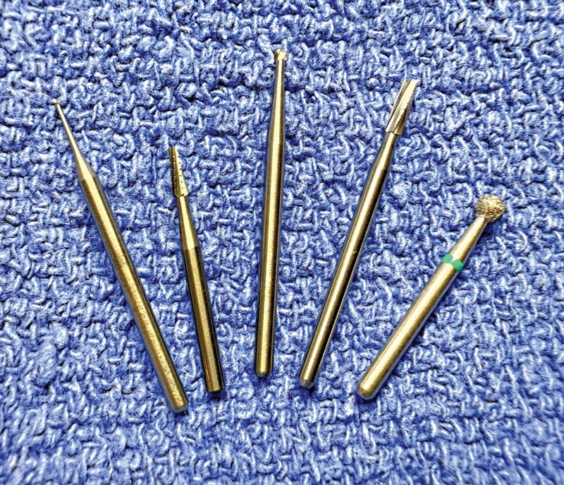
- The Periodontal Probe: Designed to measure gingival sulcus depths at multiple circumferential sites around each tooth, the probe differentiates a healthy, normal anatomic sulcus from a pathological periodontal pocket driven by attachment loss. Normal sulcal depth thresholds are remarkably tight: less than 1 mm in felines and less than 3 mm in canines. The author’s preferred instrument is the UNC-15 probe, which features clear, easy-to-read millimeter markings up to 15 mm. Beyond periodontal evaluation, the UNC-15 is exceptionally useful for measuring and documenting the precise dimensions of oral masses during oncological workups.
- The Explorer: This tactile instrument is deployed to assess structural integrity. It detects pulp exposure in fractured teeth, evaluates the gingival margin for the subtle irregularities of early feline tooth resorption, and maps out subgingival calculus deposits. Specialized tips, such as the TU #17 (or its counterpart, the Orban #17), offer delicate angles ideal for locating subtle feline tooth resorption lesions that would otherwise escape detection when using larger instruments like the classic #23 shepherd’s hook.
Official Statements and Expert Insights
To understand the operational hurdles facing general practices, we must examine the intersection of institutional training and clinical reality. As Dr. John Lewis observes:
"After 29 years in practice, I finally figured out why most veterinarians do not like dentistry. When I travel to visit veterinary colleagues at referring practices, it becomes clear most are lacking the resources they need to make surgical extractions easier. Sometimes, the problem is the lack of training because dentistry as a discipline did not have much of a foothold in their veterinary school’s curriculum."
This systemic shortfall, however, is not a permanent sentence. Dr. Lewis emphasizes that proactive post-graduate education can radically alter a clinician’s perspective:
"This is unfortunate, since dentistry is a huge part of the graduating veterinarian’s daily life. A lack of training can be addressed even after graduation by enrolling in an appropriate continuing education program. No matter what level of dental training vets have achieved, there’s always an opportunity to take it to the next level."
Shifting from diagnostics to therapeutic intervention, the choice of surgical hardware dictates procedural efficiency. High-speed and low-speed dental units form the third essential pillar of the modern operatory. Traditionally powered by compressed air, the landscape is shifting toward advanced electric motor units:
"Most units run on compressed air, but in recent years, electric units have been gaining momentum in veterinary dentistry and oral surgery. The electric unit handpiece is heavier than an air-driven handpiece, but its main benefit is the torque it can generate."
When pairing these power units with their working ends—burs—selection is critical for sectioning roots and removing alveolar bone safely:
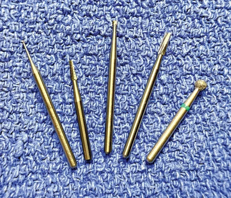
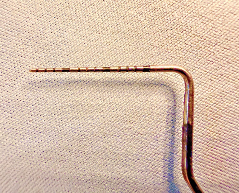
"Regardless of whether you use an air-driven or electric handpiece, having an assortment of burs is essential. This is the working end of your handpiece, and a variety of burs allows for a variety of functions… including carbide round burs, crosscut fissure burs, and diamond burs. Most carbide burs are best purchased in surgical length rather than standard length."
Future Outlook: Elevating the Standard of Care
As veterinary medicine marches further into the 21st century, client expectations and technological advancements are converging to elevate the standard of oral healthcare. Pet owners increasingly view their companion animals as family members, demanding comprehensive preventive care and pain management that matches human standards. Oral pain—long suffered silently by dogs and cats—is now recognized as a major contributor to systemic inflammation, behavioral changes, and diminished quality of life.
The future of veterinary dentistry lies in the seamless integration of high-resolution diagnostics, ergonomic instrumentation, and continuous professional development. Clinics that break free from legacy limitations—investing in state-of-the-art digital radiography, precise periodontal charting instruments, and high-torque electric surgical units—will find that procedures once dreaded as arduous chores become predictable, efficient, and clinically gratifying.
In the second installment of this series, our exploration will dive deeper into specialized extraction equipment, evaluating modern lever-action instruments, periotomes, and emerging innovations designed to streamline surgical workflows and optimize patient outcomes.
About the Author:
John Lewis, VMD, DAVDC, Fellow-AVDC OMFS, teaches at the Silo Academy Education Center and practices at Veterinary Dentistry Specialists in Chadds Ford, Pennsylvania.