
By Kendra Freeman, DVM, MS, DACVS (Large Animal/Small Animal)
Advanced Veterinary Clinical Reporting & Diagnostic Review
Executive Overview
Acute abdominal emergencies in young, large-breed canines demand immediate, methodical, and high-precision clinical intervention. Among the differential diagnoses that strike terror into the hearts of emergency clinicians and board-certified surgeons alike is gastrointestinal intussusception—a dynamic, potentially catastrophic mechanical obstruction where a segment of the intestine telescopes into an immediately adjacent segment.
This comprehensive clinical report examines a classic presentation of this surgical emergency: an 11-month-old German Shepherd Dog presenting with a stark 48-hour history of acute vomiting, profound lethargy, and severe abdominal discomfort. By analyzing this specific case presentation, veterinary professionals can re-evaluate core anatomical predilections, pathological triggers, and advanced surgical management strategies.
Beyond the immediate case study, this article investigates current veterinary literature regarding recurrence prevention, specifically evaluating the efficacy of enteral plication techniques following manual reduction. Through a rigorous journalistic and clinical lens, we explore the pathophysiological cascade of intussusception, diagnostic imaging protocols, and the critical decision-making trees that differentiate a successful recovery from catastrophic intestinal necrosis and septic peritonitis.
Detailed Chronology: Case Presentation and Diagnostic Workup
The Initial Presentation
An 11-month-old intact male German Shepherd Dog was presented to an emergency veterinary referral center with a 48-hour history of rapidly progressive clinical decline. According to the primary caretakers, the juvenile dog exhibited intermittent acute vomiting, which quickly escalated to persistent retching and the inability to retain water or food. Concurrently, the dog demonstrated marked lethargy, refusing to rise, alongside overt behavioral indicators of acute abdominal discomfort, including a hunched posture, stretching frequently, and vocalizing when the cranial and mid-abdominal quadrants were palpated.
Initial Triage and Stabilization
Upon presentation, the patient was classified as a high-priority emergency triage case. Vital parameters revealed:
- Temperature: $37.8^circtextC$ ($100.0^circtextF$)—mildly hypothermic, suggestive of systemic inflammatory response or early shock.
- Heart Rate: $140text beats per minute$—tachycardic, consistent with pain, dehydration, and hypovolemia.
- Respiratory Rate: $36text breaths per minute$—tachypneic secondary to stress and abdominal discomfort.
- Mucous Membranes: Tacky, pale pink, with a capillary refill time (CRT) prolonged to approximately 2.5 seconds.
Immediate vascular access was secured via placement of a large-bore intravenous catheter in the cephalic vein. Fluid resuscitation was initiated immediately using an isotonic crystalloid solution (Normosang-R or Lactated Ringer’s Solution) administered at an initial shock-rate bolus of $15text mL/kg$ over 15 minutes, adjusted dynamically based on perfusion parameters.
Diagnostic Imaging and Ultrasonography
With stabilization underway, diagnostic imaging was prioritized to evaluate the acute abdomen. Survey abdominal radiography revealed a marked loss of serosal detail in the mid-abdomen, coupled with several gas- and fluid-distended small intestinal loops exhibiting an abnormal stacking pattern, highly indicative of a mechanical obstruction.
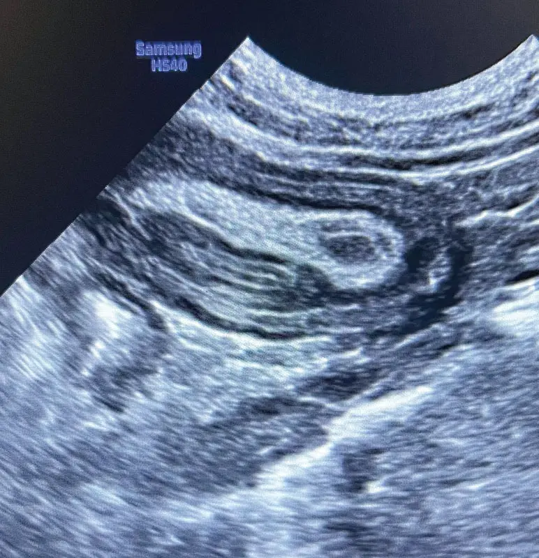
To confirm the diagnosis and assess vascular compromise, an abdominal point-of-care ultrasound (POCUS) and a comprehensive abdominal ultrasound scan were performed (referencing the clinical image provided courtesy of Dr. Amanda Rouse). The ultrasonographic evaluation revealed the classic pathognomonic sign of an intussusception: the "bull’s-eye" or "target" lesion appearance in transverse cross-section, and the "layered" or " sandwich" sign in longitudinal section.
[Transverse View: "Bull's-Eye" / "Target" Lesion]
      ( O ( O ) O )  --> Concentric rings of edematous bowel wall

[Longitudinal View: "Hay-Fork" / "Sandwich" Sign]
      ================= Outer Intussuscipiens =================
          --------------- Inner Intussusceptum ---------------
      ========================================================
The ultrasound demonstrated hypermotility and marked thickening of the bowel wall layers, localized fluid accumulation within the peritoneal cavity, and a localized loss of normal sliding motility within the affected segment. Crucially, color Doppler imaging indicated severely compromised perfusion within the innermost telescoped segment (the intussusceptum), elevating the clinical urgency for immediate exploratory laparotomy.
Clinical Challenges & Core Knowledge Evaluation
To test and reinforce advanced clinical competence in managing gastrointestinal emergencies, veterinary practitioners must evaluate three pivotal questions regarding the anatomy, etiology, and surgical management of canine intussusception.
1) In dogs, the most common location for an intussusception is:
- a. Ileocolic junction
- b. Jejunojejunal
- c. Jejunoileal
- d. Coloncolic
Clinical Discussion:
The correct answer is a. Ileocolic junction.
While intussusceptions can theoretically occur at any point along the continuous tract of the alimentary canal, anatomical and physiological factors heavily favor the ileocolic junction in dogs. Young animals—particularly those under one year of age, such as our 11-month-old German Shepherd—are disproportionately affected. The abrupt transition in luminal diameter, differential motility patterns, and structural fixation points between the terminal ileum, cecum, and proximal colon create a dynamic fulcrum where telescoping frequently occurs.
2) The most common underlying cause of canine intussusceptions is:
- a. Parasitic
- b. A foreign material
- c. Neoplasia
- d. Idiopathic
Clinical Discussion:
The correct answer is d. Idiopathic (though frequently secondary to an underlying enteritis or motility disorder, a vast majority of cases in young dogs have no identifiable structural mass or foreign body acting as a lead point).
In pediatric and adolescent canine populations, severe parasitism (e.g., Ancylostoma caninum or Toxocara canis), acute viral enteritis (such as Canine Parvovirus), dietary indiscretion, or functional dysmotility frequently trigger localized hyperperistalsis. This chaotic, uncoordinated hypermotility allows a normal segment of bowel to telescope into an adjacent, more relaxed segment. Conversely, older dogs presenting with intussusception are significantly more likely to harbor an underlying structural "lead point," such as linear foreign bodies, intestinal neoplasia (adenocarcinoma, lymphoma, leiomyosarcoma), or polyps.
3) According to Larose et al., enteral plication after manual reduction of an intussusception prevents recurrence of the intussusception.
- a. True
- b. False
Clinical Discussion:
The correct answer is b. False.
Enteral plication (mesenteric plication or Noble plication), historically debated as a method to prevent recurrent intussusceptions or chronic small intestinal volvulus by fixing loops of the small intestine in ordered, gentle folds, does not guarantee absolute prevention and carries substantial iatrogenic risks. Contemporary veterinary surgical literature, including evaluations by Larose and other surgical researchers, highlights that enteral plication is associated with significant potential complications, including needle-tract leakage, focal intestinal necrosis, persistent localized pain, adhesion formation, and subsequent mechanical obstructions. Consequently, modern board-certified veterinary surgeons (DACVS) generally advocate for targeted resection and anastomosis (R&A) of compromised bowel segments combined with correction of underlying primary enteropathies, rather than routine prophylactic plication.
Supporting Context & Metrics: Epidemiology and Pathophysiology
Epidemiological Trends
Gastrointestinal intussusception is predominantly a disease of the young. Retrospective veterinary epidemiological data indicate that approximately 70% of canine intussusception cases occur in patients less than one year of age. Large and giant-breed dogs—including German Shepherd Dogs, Labrador Retrievers, and Poodles—are overrepresented in surgical caseloads. This breed predilection may be tied to anatomical variations in mesenteric attachment lengths, higher rates of juvenile parasitic infections, and dietary indiscretion habits common in adolescent working and sporting breeds.
The Pathophysiological Cascade
The formation of an intussusception sets off a rapid and merciless pathological cascade:
- Initial Telescoping: The proximal segment (intussusceptum) invaginates into the distal lumen (intussuscipiens).
- Venous Obstruction: As the bowel walls telescope into one another, the mesentery belonging to the intussusceptum is pulled along and compressed. Because veins possess lower intraluminal pressure and thinner walls than arteries, venous outflow is obstructed long before arterial inflow is compromised.
- Congestion and Edema: Severe venous and lymphatic congestion ensues, causing the trapped intestinal walls to become massively edematous, hemorrhagic, and friable.
- Ischemia and Necrosis: As interstitial pressure rises and arterial flow eventually fails, full-thickness ischemic necrosis develops within hours.
- Translocation and Peritonitis: Compromise of mucosal barrier integrity permits bacterial translocation across the bowel wall, rapidly precipitating septic peritonitis, systemic inflammatory response syndrome (SIRS), and, if untreated, death.
[Invagination]
      │
      ▼
[Mesenteric Traction & Compression]
      │
      ▼
[Venous Outflow Obstruction (Early)]
      │
      ▼
[Severe Edema, Hemorrhage, & Congestion]
      │
      ▼
[Arterial Inflow Compromise (Late)]
      │
      ▼
[Ischemic Necrosis & Bacterial Translocation]
      │
      ▼
[Septic Peritonitis & Circulatory Collapse]
Official Statements and Surgical Perspectives
Insights from Veterinary Surgeons
Dr. Amanda Rouse, whose diagnostic imaging provided the foundational ultrasound reference for this case, emphasizes the critical nature of early ultrasonographic screening.
"When evaluating an adolescent large-breed dog exhibiting acute vomiting and painful cranial abdominal palpation, time is quite literally tissue," notes Dr. Rouse. "Plain radiography offers vital clues regarding mechanical obstruction, but ultrasonography remains the gold standard for visualizing the dynamic wall layers of an intussusception. Identifying loss of perfusion via Doppler interrogation before the patient spirals into irreversible septic shock alters the entire surgical trajectory."
Adding to this perspective, surgical specialists emphasize that exploratory laparotomy must be approached with meticulous tissue handling. Once the abdomen is entered via a standard ventral midline celiotomy, the affected segment must be carefully exteriorized and packed off with warm, saline-soaked laparotomy sponges to prevent peritoneal contamination.
Surgical Decision-Making: Reduction vs. Resection
The attending surgical team faces a crucial binary decision upon exteriorizing the intussusception:
- Manual Reduction: Attempting to gently push the intussusceptum out of the intussuscipiens using retrograde milking motions (never pulling, as pulling increases the risk of iatrogenic tearing).
- En Bloc Resection and Anastomosis (R&A): Directly resecting the entire affected, non-viable, or severely compromised intestinal complex.
Board-certified large and small animal surgeons stress that manual reduction should only be attempted if the bowel wall retains healthy serosal color, normal peristaltic response, palpable pulses in the mesenteric arcades, and a lack of overt friability. If the tissue appears dusky, dark purple, black, or green, or if it demonstrates antemortem perforation, immediate en bloc resection and end-to-end anastomosis are mandatory. Attempting to reduce a necrotic intussusception releases massive quantities of endotoxins and inflammatory mediators directly into the systemic circulation ("reperfusion injury"), while leaving necrotic tissue in situ invites catastrophic dehiscence and fatal peritonitis.
Future Outlook and Clinical Recommendations
As veterinary medicine continues to advance, the management of acute abdominal emergencies like canine intussusception relies heavily on early intervention, high-resolution diagnostic imaging, and precise surgical technique.
Key Recommendations for Clinical Practice:
- Low Threshold for Ultrasound: Emergency clinicians presented with vomiting juvenile dogs—particularly large-breed males showing abdominal pain—should maintain a high index of suspicion for intussusception and perform immediate abdominal ultrasonography.
- Aggressive Fluid Resuscitation: Preoperative stabilization with balanced isotonic crystalloids, targeted colloid therapy where indicated, and appropriate multimodal analgesia (including constant rate infusions of opioids such as fentanyl or ketamine) are non-negotiable prerequisites for safe general anesthesia.
- Prudent Surgical Conservatism: Surgeons must prioritize patient safety by maintaining a low threshold for intestinal resection and anastomosis over risky manual reductions of compromised, ischemic bowel.
- Postoperative Care & Monitoring: Intensive postoperative care must include close monitoring for signs of ileus, continued fluid therapy, electrolyte balance management, and vigilant observation for clinical indicators of septic peritonitis or anastomotic dehiscence.
Through strict adherence to these evidence-based protocols, veterinary practitioners can dramatically improve survival rates and long-term outcomes for canine patients afflicted with this complex and life-threatening surgical emergency.