
Executive Overview
Deep within the structural architecture of the modern human brain lies a profound evolutionary relic: the pineal gland. Long recognized by neuroscientists as the body’s master regulator of circadian rhythms and sleep-wake cycles, this pinecone-shaped endocrine organ carries an ancestry far stranger than previously imagined. According to groundbreaking research spearheaded by evolutionary biologists at Lund University and the University of Sussex, humanity and all other living vertebrates trace their lineage back to a tiny, worm-like marine organism that navigated the primordial oceans nearly 600 million years ago with a single, central eye perched atop its head.
This startling revelation—published recently in a comprehensive comparative analysis of sensory biology—turns conventional evolutionary theory upside down. For decades, standard evolutionary models assumed that the paired, image-forming eyes of vertebrates evolved in a linear progression from simple, multi-ocular ancestral setups. However, the new data suggests a far more circuitous and bizarre historical detour. Our distant forebears abandoned an active, mobile lifestyle to adopt a sedentary, filter-feeding existence. In doing so, they cast off their paired lateral eyes as evolutionary dead weight, relying instead on a rudimentary, central median eye to sense the crude shifts between day and night.
Millions of years later, as their descendants returned to an active, swimming lifestyle, evolutionary pressures forced a radical reinvention. Rather than growing a new set of eyes from scratch or repairing the old ones, nature repurposed the cells of that ancient median eye, giving rise to the complex, paired optical systems we possess today. Furthermore, this unusual developmental trajectory explains a foundational anatomical mystery that has puzzled biologists for generations: why vertebrate eyes, featuring retinas that sprout directly from embryonic brain tissue, are fundamentally inverted compared to the skin-derived, camera-like eyes of cephalopods like squids or arthropods like insects.
As we explore the intricate mechanics of this discovery, we uncover not only the origins of our sight, but also the deep biological heritage that links human sleep architecture directly to the cyclopean gaze of a Precambrian worm.
Detailed Chronology: From Precambrian Seas to the Vertebrate Brain
To understand how a single-eyed marine organism laid the groundwork for human vision and neurological function, science must journey back approximately 600 million years to the late Precambrian era. This was a critical juncture in Earth’s history, predating the sudden explosion of complex animal life known as the Cambrian explosion. The oceans were shifting chemically, and early multicellular life forms were experimenting with novel body plans, metabolic pathways, and sensory organs.
Phase 1: The First Split and the Loss of Lateral Vision
Before our lineage settled on a single-eyed configuration, the very earliest chordate and vertebrate ancestors possessed rudimentary light-sensitive cell patches—effectively, paired visual inputs. In the open water, these paired detectors provided a distinct survival advantage, allowing mobile organisms to judge direction, track passing shadows, and calculate distance relative to moving hazards or potential food sources.
However, life is ruthlessly economical. As one specific branch of these ancient organisms transitioned to a sessile, stationary lifestyle—anchoring themselves to the ocean floor and filtering passing plankton from the seawater—the high metabolic and developmental costs of maintaining complex paired eyes became a liability. Without the need to hunt, evade active predators, or navigate complex topological terrain, the genes governing lateral eye development were sidelined. Over countless generations, these paired eyes faded away entirely through the gradual accumulation of neutral mutations and natural selection against unnecessary biological overhead.
Phase 2: The Reign of the Median Eye
While the lateral eyes vanished, a solitary cluster of photoreceptive cells situated directly on the dorsal midline of the creature’s head remained intact. This median, or "cyclopean," eye did not produce high-resolution, sharp images in the modern sense. Instead, it operated as a crude environmental sensor.
For a stationary filter-feeder, knowing the precise orientation of the water column was a matter of life and death. The median eye provided this vital orientation data by detecting the vector of sunlight piercing the water’s surface. It allowed the organism to distinguish day from night and determine upward versus downward, guiding its metabolic processes and anchoring posture relative to the photic zone. This single-eyed configuration sustained the lineage through vast expanses of geological time, proving that minimalism can be an effective survival strategy when an animal’s ecological niche requires little movement.
Phase 3: The Great Return to Mobility and the Invention of Paired Eyes
The evolutionary stasis of the cyclopean phase was eventually broken. Driven by shifting ecological pressures, competition for resources, or new environmental niches, the descendants of these sessile filter-feeders abandoned their anchored existence and returned to the water column as active swimmers.
A mobile lifestyle immediately resurrected the need for sophisticated spatial awareness. Swimming animals cannot survive long without the ability to detect predators, locate moving prey, and steer clear of physical obstacles. Nature was faced with an evolutionary engineering challenge: how to build a high-performance visual system for a fast-moving body plan.
Rather than resurrecting the long-lost lateral eye pathways from earlier in the lineage, evolution engineered a remarkable workaround. It co-opted portions of the existing median visual structure. Through a series of complex genetic shifts, cells from this central region migrated, multiplied, and specialized, ultimately giving rise to a brand-new pair of lateral, image-forming eyes.
This unexpected genealogical link clarifies why vertebrate retinas develop inward-out—with light having to pass through layers of neurons and blood vessels before hitting the photoreceptor cells—while invertebrate eyes developed from the outer ectoderm (skin). Because vertebrate eyes originated from central brain tissue, their neural wiring and retinas carry the unmistakable developmental fingerprints of their neurological birthplace.
Supporting Context & Metrics: The Anatomy of an Evolutionary Relic
To fully appreciate the weight of this discovery, it is necessary to examine the physical and biological evidence that allowed researchers at Lund University and the University of Sussex to reconstruct this ancient timeline. The team’s conclusions were not drawn from the fossil record alone—soft-bodied Precambrian organisms rarely fossilize in ways that preserve delicate neural or retinal architecture. Instead, the researchers relied on modern comparative developmental biology, gene-expression mapping, and comparative neuroanatomy.
Comparative Metrics of Eye Development Across Taxa
| Animal Group | Primary Evolutionary Origin of Eye Tissue | Associated Visual Structures | Evolutionary Strategy |
|---|---|---|---|
| Vertebrates (Humans, Fish, Birds) | Neuroectoderm (Outgrowth of embryonic brain tissue) | Paired lateral eyes, inverted retina, neural circuits in retina | Repurposed median eye structure following secondary mobility |
| Arthropods (Insects, Crustaceans) | Surface Ectoderm (Infolding of skin tissue) | Compound eyes, ocelli | Direct linear evolution of external light-detecting skin patches |
| Molluscs (Squid, Octopus) | Surface Ectoderm (Thickening and depression of skin) | Camera-type eyes with unverted retinas | Convergent evolution optimized for active predation |
As illustrated above, vertebrates stand apart in the animal kingdom due to the cerebral origin of their optical apparatus. The retina is, quite literally, a piece of the brain pushed outward during embryonic development. This structural peculiarity creates an evolutionary paradox: why would an optical sensor develop inside the neural tube and face inward, rather than forming directly on the exterior surface where light hits unhindered? The cyclopean ancestor theory provides the first cohesive, logical answer. When the central median eye was expanded and split to form new lateral eyes, those eyes retained the genetic and developmental programming of the brain tissue from which they originally sprouted.
The Pineal Gland: Living History in the Human Skull
Perhaps the most fascinating manifestation of this theory is the persistence of the median eye’s structural remnants within the modern human brain. The pineal gland—often romantically referred to by philosophers like René Descartes as the "principal seat of the soul"—is a tiny, reddish-grey endocrine gland located in the epithalamus, nestled between the two cerebral hemispheres.
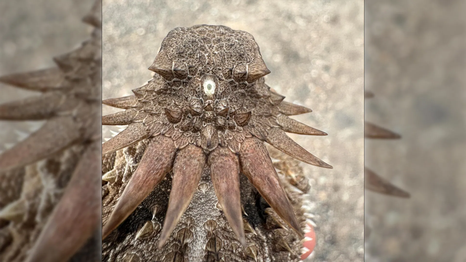
While humans do not use the pineal gland to see images, its cellular composition reveals its ancient visual heritage. The gland contains pinealocytes, specialized cells that evolved from photoreceptors. In many lower vertebrates—such as certain fish, amphibians, and reptiles—the pineal organ (sometimes called the "parietal eye" or "third eye") sits directly beneath a gap in the skull, covered only by skin and scales, and functions as a direct light sensor that helps synchronize seasonal behaviors and physiological thermoregulation.
In mammals, the pineal gland has lost its direct photoreceptive function, delegating the task of light detection to the specialized retinal ganglion cells in our paired eyes. However, the downstream neural pathways remain remarkably conserved:
- Light Detection: Photons strike the retina, activating specialized melanopsin-containing retinal ganglion cells.
- Neural Transmission: Signals travel via the retinohypothalamic tract to the suprachiasmatic nucleus (SCN) of the hypothalamus, the master biological clock.
- Endocrine Response: The SCN relays inhibitory or stimulatory signals through a complex autonomic pathway to the superior cervical ganglion, which ultimately innervates the pineal gland.
- Hormonal Regulation: In the absence of light (nighttime), the pineal gland synthesizes and secretes melatonin, the neurohormone responsible for signaling to the rest of the body that it is time to rest, lower core body temperature, and initiate restorative sleep cycles.
Thus, every time a human being experiences jet lag, drifts off to sleep under the quiet of night, or feels the seasonal lethargy of winter, they are engaging a biochemical cascade mediated by an organ that once served as the sole, cyclopean gaze of a Precambrian worm.
Official Statements and Expert Analysis
The implications of this research extend far beyond academic journals, challenging fundamental assumptions about how complex biological systems evolve. The study’s lead researchers have emphasized both the shock of the findings and the clarity they bring to longstanding anatomical anomalies.
"The results are a surprise. They turn our understanding of the evolution of the eye and the brain upside down,"
— Dan-E Nilsson, Professor Emeritus in Sensory Biology at Lund University.
Professor Nilsson, a world-renowned authority on the evolution of eyes, points out that tracing the lineage of visual components requires looking past superficial similarities and examining deep genetic and developmental blueprints. For decades, evolutionary biologists debated whether vertebrate eyes and invertebrate eyes shared a single point of origin or represented a textbook example of convergent evolution—where similar traits evolve independently in unrelated lineages. This new research refines that debate by showing that while the functional outcome (paired, image-forming eyes) is indeed convergent, the raw materials used by vertebrates to build their eyes were fundamentally recycled from an internal, single-eyed organ.
Furthermore, Nilsson highlights how this framework solves a major mystery regarding the neural architecture of our visual system:
"Now we finally understand why the eyes of vertebrates differ so radically from the eyes of all other animal groups, such as insects and squid. The film of our eyes—the retina—developed from the brain, whereas the eyes of insects and squid originate in the skin on the sides of the head… For the first time, we now also understand the origin of the neural circuits that analyze the image in our retina."
Independent neurobiologists and evolutionary theorists have praised the study for bridging the gap between molecular embryology and macro-evolution. By demonstrating how ancient neural circuits—originally designed to process simple light/dark distinctions in a primitive median eye—were scaled up, duplicated, and co-opted to handle high-resolution visual processing, the research provides a mechanistic model for how complex biological novelties emerge from simpler, repurposed precursors.
Future Outlook: What the Cyclops Ancestor Teaches Us About Modern Science
As evolutionary biology enters an era defined by advanced genomic sequencing, single-cell transcriptomics, and high-resolution imaging, the discoveries made by the Lund and Sussex research teams open exhilarating new avenues for scientific inquiry.
1. Rethinking Evolutionary Plasticity
The revelation that vertebrates underwent a "cyclopean detour"—sacrificing complex paired eyes only to reinvent them later from central brain tissue—underscores the astonishing plasticity of evolutionary processes. Evolution is not a straight, forward-marching ladder of progressive improvement; it is a messy, opportunistic tinkerer. Traits are lost, reclaimed, downsized, and radically repurposed when ecological demands shift. This insight encourages modern synthetic biologists and bioengineers to look at biological systems not as fixed, rigid architectures, but as dynamic networks capable of radical functional reinvention.
2. Clinical Implications for Sleep and Neurological Research
Understanding the deep evolutionary origins of the pineal gland and its connection to light-sensing pathways may also offer fresh perspectives in clinical medicine. Circadian rhythm disorders, seasonal affective disorder (SAD), and age-related disruptions in melatonin production are major public health challenges in our modern, screen-lit society. By mapping the exact neural and molecular conduits that link our visual system to our internal endocrine regulators, researchers can design more targeted phototherapy interventions, pharmacological treatments for insomnia, and lifestyle frameworks that respect our ancient biological wiring.
3. The Ongoing Quest into the Tree of Life
Ultimately, the story of our one-eyed Precambrian ancestor serves as a humbling reminder of our deep connections to the history of planet Earth. As marine biologists and paleontologists continue to unearth microfossils and decode the genomes of basal chordates like amphioxus and tunicates, the picture of our earliest origins will only sharpen.
The next time you look up at the stars, or feel the heavy pull of sleep at the end of a long day, consider the journey of your ancestors. From a tiny, worm-like creature drifting in the dark, light-dappled oceans of a 600-million-year-old world, peering upward with a single, unblinking eye, to the complex, conscious minds we possess today—our capacity to perceive the universe is an inheritance forged through adaptation, loss, and an extraordinary evolutionary reinvention.