
Executive Overview
Deep within the intricate architecture of the human brain rests a tiny, pinecone-shaped structure known as the pineal gland. Long recognized for its role in synthesizing melatonin and regulating our circadian rhythms, this endocrine organ has captivated philosophers and scientists for centuries—most famously dubbed "the principal seat of the soul" by the 17th-century French philosopher René Descartes. However, groundbreaking new evolutionary research reveals that the pineal gland is far more than a chemical regulator of sleep. According to a collaborative study conducted by evolutionary biologists and sensory specialists at Lund University and the University of Sussex, this deep-brain structure is the direct living descendant of a single, centrally located eye that adorned the top of the head of our earliest vertebrate ancestors some 600 million years ago.
This paradigm-shifting discovery challenges fundamental assumptions regarding the timeline and mechanics of ocular and neurological development. For generations, evolutionary biology operated under the general consensus that the vertebrate visual system evolved along a relatively linear path from simple light-sensitive patches to complex, image-forming paired eyes. The new findings, however, reveal a bizarre and unexpected evolutionary detour: long before our ancestors swam the ancient seas with two sophisticated cameras for eyes, they passed through a "cyclopean" phase. During this period, an ancestral worm-like organism abandoned its paired eyes, relied entirely on a single median eye positioned dead-center on its head, and subsequently rebuilt an entirely new set of paired eyes from that central neural foundation.
The implications of this research extend far beyond mere biological trivia. By tracing the lineage of the vertebrate eye back to a single median organ, scientists have finally resolved a long-standing anatomical puzzle: why vertebrate eyes are structurally inverted compared to the eyes of invertebrates like insects and cephalopods. While an insect’s eye develops outward from the ectoderm—the embryonic skin—the vertebrate retina is fundamentally an extension of the central nervous system, bulging outward from the embryonic brain.
This authoritative investigative report explores the monumental findings of the Lund and Sussex research teams, detailing the chronology of this prehistoric evolutionary pivot, analyzing the structural mechanics that separate vertebrate vision from the rest of the animal kingdom, examining official statements from the leading architects of the study, and casting a glance toward how this ancient heritage continues to dictate the rhythm of human life today.
Detailed Chronology: The 600-Million-Year Journey from Worm to Vertebrate
To understand how humanity inherited a neurological remnant of a mythological monster, science must peer back into the Precambrian and early Cambrian epochs, approximately 600 million years ago. The world was radically different; continents were locked in primordial configurations, and complex multicellular life was undergoing a sweeping diversification.
Phase I: The Primal Pair and the Stationary Shift
According to the fossil record and comparative genomic analyses, the common ancestor of all living vertebrates was a diminutive, soft-bodied, worm-like organism. In the earliest chapters of its lineage, this creature likely possessed paired, rudimentary light-sensitive spots—simple predecessors to eyes that aided in detecting the looming shadows of predators or navigating the ambient gradients of ancient photic zones.
However, evolutionary pressures are rarely static. As this worm-like organism adapted to a more sedentary, benthic lifestyle, anchoring itself to the sea floor to filter plankton from passing ocean currents, the energetic and ecological costs of maintaining complex, mobile sensory structures shifted. For a creature that spent most of its life anchored in one place, wide-ranging stereoscopic or paired vision offered diminishing returns.
"We don’t know whether the paired eyes in our branch of the evolutionary tree were just light-sensitive cells or simple image-forming eyes," explains Dan-E Nilsson, professor emeritus in sensory biology at Lund University and a leading voice in the new study. "We only know that the organism later lost them."
Phase II: The Cyclopean Era
With the gradual regression and eventual loss of the paired lateral eyes, the organism did not become entirely blind. A cluster of specialized, light-sensitive cells persisted in the direct center of the dorsal surface of its head. Over countless generations, natural selection honed this cluster into a functional, singular median eye.
Positioned atop the head like a tiny, biological periscope, this "cyclopean" organ served vital survival functions. It did not project high-definition cinematic images of the Precambrian world, but it excelled at basic phototaxis—distinguishing between the blinding safety of the sunlit surface and the dark, perilous depths, while assisting the organism in spatial orientation. For millions of years, this single median eye was the sole optical window through which the lineage of all future fish, amphibians, reptiles, birds, and mammals perceived the light of day.
Phase III: The Swimming Renaissance and the Rebuilding of Vision
The evolutionary trajectory of this lineage shifted once more when the organisms abandoned their stationary, filter-feeding habits and returned to an active, swimming lifestyle. Mobility introduced entirely new selective pressures. Navigating open waters, hunting elusive prey, and evading mobile predators demanded sophisticated spatial awareness and high-resolution environmental mapping.
Rather than reinventing ocular structures from scratch or magically regenerating the long-lost lateral eyes of their distant ancestors, these swimming descendants did something extraordinary. They co-opted the existing neural machinery and light-sensitive cell lines of the median eye. Through radical developmental transformations, parts of this original central eye structure expanded, bifurcated, and adapted to give rise to a brand-new set of paired, image-forming eyes.
This sweeping architectural reorganization bypassed the standard developmental pathways seen in other phyla, locking vertebrates into a fundamentally distinct blueprint for sight—one where the light-gathering screen of the eye is wired directly into the processing units of the brain from the very beginning of embryonic development.
Supporting Context & Metrics: Unraveling the Vertebrate Blueprint
The revelation that vertebrate eyes stem from a single median organ resolves one of comparative anatomy’s most stubborn riddles: the profound developmental dichotomy between vertebrate eyes and those of successful invertebrate groups like insects, spiders, and cephalopods (such as squid and octopuses).
The Inverted Retina Paradox
For over a century, evolutionary developmental biologists have marveled at the "inverted" nature of the vertebrate retina. In a human eye, light must pass through a dense web of blood vessels, ganglion cells, and bipolar neurons before it finally strikes the light-sensitive rods and cones tucked away at the very back of the retinal wall. To an engineer, this design appears counterintuitive—like placing the film of a camera behind a tangle of wires and wiring harnesses.
In contrast, the eyes of an octopus or a housefly are constructed with direct-illumination retinas. Their photoreceptor cells face forward, meeting incoming light head-on without obstruction.
The research from Lund and Sussex explains why this striking structural variance exists:
[Invertebrate Lineage (Insects/Squid)]
--> Develops outward from surface ectoderm (skin tissue)
--> Direct-illumination retina (Photoreceptors face forward)

[Vertebrate Lineage (Humans/All Backboned Animals)]
--> Develops as an extension of the central nervous system (brain tissue)
--> Inverted retina (Photoreceptors face backward behind neural layers)
    * Rooted in the ancient median eye of a 600-million-year-old ancestor
Because vertebrate eyes were rebuilt from a central median eye—which was fundamentally an outpost of neural tissue located on top of the head—the resulting visual apparatus retained its cerebral identity. The retina is, quite literally, a specialized piece of the brain pushed outward toward the skull. Insect and squid eyes, meanwhile, evolved independently from the surface ectoderm (skin tissue) on the sides of the head. This divergent embryonic origin explains the deep structural gulf separating our vision from that of the invertebrate world.
Cellular and Genetic Signatures
To arrive at these conclusions, the international research team did not rely solely on theoretical models or the fossil record. They performed rigorous comparative analyses of light-sensitive proteins (opsins), neural circuits, and embryological growth factors across a vast spectrum of living and extinct animal lineages.
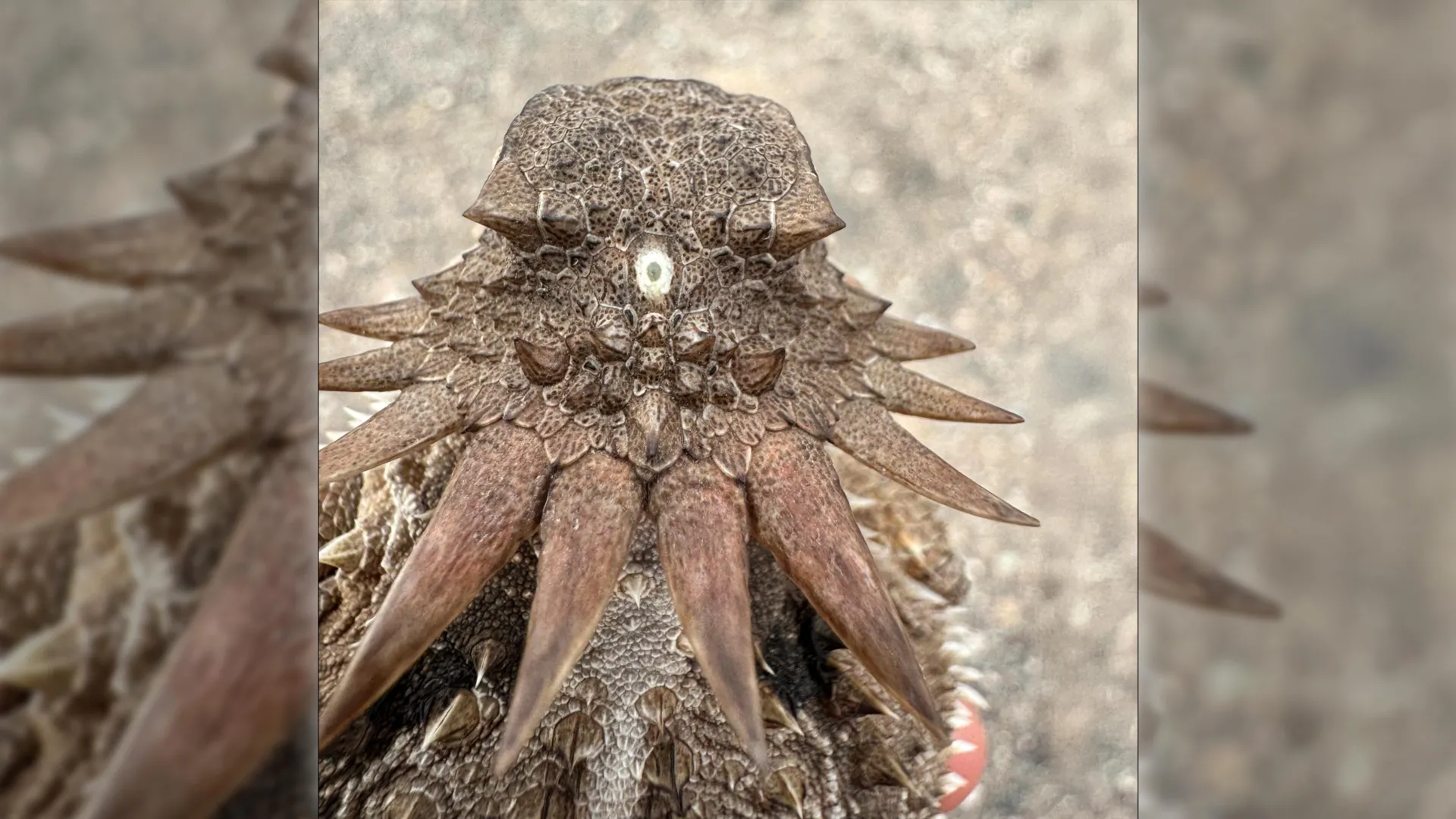
By mapping the genetic pathways that govern the development of the pineal gland in modern vertebrates and comparing them with the pineal eyes (parietal eyes or "third eyes") still found in living "living fossils" like the tuatara (Sphenodon punctatus)—a reptile native to New Zealand—scientists mapped a clear trail of cellular homology. The biochemical machinery responsible for melatonin production in the human pineal gland shares direct molecular ancestry with the light-transducing pigments that once helped our Precambrian ancestors track the cycles of the sun.
Official Statements and Expert Analysis
The publication of these findings has sent ripples through the global scientific community, prompting profound reflections on how human anatomy reflects deep-time evolutionary history.
"The results are a surprise," says Dan-E Nilsson, professor emeritus in sensory biology at Lund University, whose decades of research into eye evolution have culminated in this watershed moment. "They turn our understanding of the evolution of the eye and the brain upside down."
Nilsson emphasizes that the journey from a sessile, worm-like filter feeder to active, intelligent vertebrates required an extraordinarily improbable sequence of biological events—a detour that ultimately defined the sensory reality of every mammal, bird, reptile, amphibian, and fish on Earth.
"For the first time, we now also understand the origin of the neural circuits that analyze the image in our retina," Nilsson notes, pointing out that tracing the visual system back to the median eye clarifies how the brain’s visual cortex and retinal layers established their sophisticated communication networks.
Other members of the collaborative research team from the University of Sussex highlighted the philosophical and physiological weight of the discovery, particularly regarding the pineal gland. Far from being a modern evolutionary afterthought or a vestigial organ with fading utility, the pineal gland stands as an ancient monument to our biological origins.
"It’s mind-boggling that our pineal gland’s ability to regulate our sleep according to light stems from the cyclopean median eye of a distant ancestor 600 million years ago," Nilsson concludes. Every time darkness falls, triggering the pineal gland to flood our bloodstream with melatonin to prepare the body for rest, we are participating in a 600-million-year-old biological ritual initiated by a tiny, one-eyed creature filtering plankton in the primeval oceans.
Future Outlook: What the "Cyclops" Ancestor Means for Modern Science
As the dust settles on this groundbreaking study, evolutionary biologists, neuroscientists, and medical researchers are already looking toward the horizon to determine how these insights will shape future investigations.
1. Advanced Evolutionary Developmental Biology (Evo-Devo)
The confirmation that vertebrate eyes underwent a structural rebuild following the loss of paired lateral eyes provides a powerful new framework for studying genomic plasticity. Researchers aim to pinpoint the specific master regulatory genes that enabled the median eye to expand and generate complex paired structures during the Cambrian explosion. Understanding how ancient genetic toolkits permitted such radical anatomical rewiring could offer profound clues into tissue regeneration and stem cell engineering in modern medicine.
2. Chronobiology and Sleep Therapeutics
Given that the human pineal gland retains its ancestral sensitivity to light—albeit mediated indirectly through the retinas and the suprachiasmatic nucleus rather than direct cranial exposure—deeper insights into its evolutionary roots could revolutionize chronobiology. Understanding the precise biochemical evolution of melatonin synthesis pathways may lead to novel therapeutic interventions for sleep disorders, seasonal affective disorder (SAD), and circadian disruption caused by modern shift work and blue-light exposure.
3. Redefining Ocular Pathology
By understanding that the vertebrate retina is fundamentally an extension of the brain, ophthalmologists and neuro-ophthalmologists gain a clearer conceptual model of ocular diseases. Conditions that bridge the eye and the brain—such as optic neuritis, glaucoma, and certain retinal degenerations—can be more accurately understood as disorders affecting an exposed, specialized outpost of the central nervous system.
Conclusion
Humanity’s place in the natural world is defined by endless layers of heritage, stretching back through epochs of dramatic transformation. The revelation that we share our deepest ocular lineage with a tiny, one-eyed, worm-like creature from the dawn of complex life humbling reminds us of the winding, unpredictable paths of evolution. From a single median eye perched atop a Precambrian head to the complex binocular vision that reads these words, our eyes—and the quiet, rhythm-keeping gland hidden deep within our brains—remain living testaments to an extraordinary evolutionary odyssey.