
Executive Overview
For generations, mammalian embryology and molecular genetics operated under a foundational assumption: that the DNA packed inside a newly fertilized egg was essentially a structural "blank slate." Conventional wisdom dictated that the maternal and paternal genomes coalesced into a loose, disorganized, and chaotic tangle upon fertilization. In this historical view, the nascent genome remained an unformatted ball of yarn, devoid of distinct spatial organization, until it experienced what geneticists refer to as "awakening"—the moment the embryo begins to actively transcribe its own genes, a monumental milestone known as Zygotic Genome Activation (ZGA). Until that genetic switch was finally thrown, the prevailing dogma held that spatial architecture simply did not matter.
A pair of landmark studies published in Nature Genetics and Nature Cell Biology has systematically dismantled this decades-old paradigm. Spearheaded by Professor Juanma Vaquerizas and his research team, alongside a collaborative companion project led by Professor Ulrike Kutay at ETH Zürich in Switzerland, these investigations reveal that the genome is far from chaotic at the dawn of life. Instead, it exhibits a staggering, highly sophisticated level of 3D structural organization long before the first embryonic genes are ever transcribed.
This architectural marvel was uncovered through the deployment of a revolutionary, high-sensitivity mapping technology dubbed Pico-C. Operating with a resolution and sample-efficiency previously thought impossible, Pico-C has allowed scientists to peer into the microscopic interior of single, newly fertilized embryos and observe an elaborate 3D scaffolding taking shape. This spatial framework is not merely a passive byproduct of nuclear packaging; it is an active, deterministic blueprint. The precise spatial folding of DNA dictates which genetic sequences will eventually be toggled on, when they will be expressed, and how cells will differentiate.
Furthermore, the implications of this research extend far beyond the opening hours of embryogenesis. In companion studies involving human cellular models, researchers demonstrated that when these 3D structural anchors fail, the consequences are catastrophic. Human cells misinterpret the structural collapse as a viral invasion, mistakenly triggering a severe innate immune response that can result in chronic inflammation, cellular dysfunction, and disease. By wedding high-resolution developmental biology with human pathology, these findings establish 3D genome architecture as a critical pillar of both life’s beginning and its ongoing cellular defense.
Detailed Chronology: From Biological Dogma to the Pico-C Breakthrough
To fully appreciate the magnitude of the recent discoveries made by Vaquerizas, lead author Noura Maziak, and their international colleagues, one must trace the historical trajectory of how geneticists have studied three-dimensional chromatin folding.
The Era of the Structural "Blank Slate"
For decades, the technical limitations of molecular biology constrained how scientists could investigate nuclear architecture. Techniques like Chromosome Conformation Capture (Hi-C) and its derivatives revolutionized the field by allowing researchers to map the physical proximity of different DNA segments within the nucleus. However, these traditional methods suffered from a major constraint: they required massive amounts of starting biological material—often millions of cells—to generate reliable maps of genome folding.
Consequently, studying the earliest moments of life, when an embryo consists of just a single cell or a small handful of dividing cells, was virtually impossible. Because researchers could not directly measure chromatin folding in these ultra-low-input settings, they relied on indirect biochemical assays and microscopic observations. These indirect methods suggested that the chromatin in newly fertilized zygotes was uniformly dispersed, lacking the complex topological domains (TADs) and looping structures observed in somatic cells. Thus, the "blank slate" hypothesis was born: a conceptual framework where the early zygotic genome waited in a quiescent, unstructured state until Zygotic Genome Activation forced it to organize.
The Development of Pico-C Technology
Recognizing that bulk-cell methods were blinding science to the critical events of early embryogenesis, Vaquerizas and his team set out to engineer a technology sensitive enough to capture the 3D architecture of ultra-small biological samples. The result of their methodological innovation is Pico-C, a high-sensitivity, high-resolution mapping protocol designed to interrogate chromatin conformation with roughly ten times less input material than standard techniques.
Pico-C achieves its remarkable efficiency by optimizing the capture and ligation steps of chromosomal interactions, minimizing sample loss, and maximizing the signal-to-noise ratio. This technological leap essentially handed geneticists a molecular microscope capable of resolving individual folds and loops inside microscopic developmental stages that were previously invisible to structural genomics.
Mapping the Fruit Fly Embryo
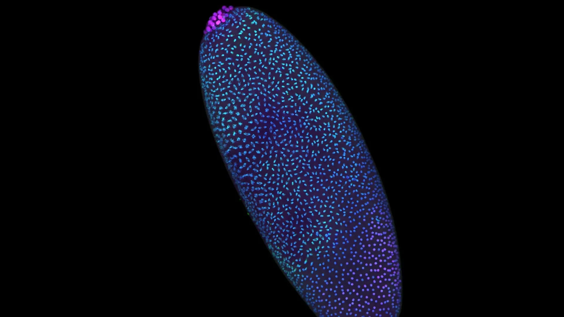
To test the capabilities of Pico-C, the researchers turned to a classical and profoundly reliable model organism in genetics: the fruit fly (Drosophila melanogaster). During the initial hours following fertilization, the fruit fly embryo undergoes a remarkably rapid and synchronized series of nuclear divisions without intervening cell divisions, producing thousands of nuclei in a matter of hours. This accelerated developmental timeline makes Drosophila an ideal system for capturing the rapid, dynamic transitions of genome reorganization.
Using Pico-C, the team mapped the 3D arrangement of the fruit fly genome across these critical early hours. The results stunned the laboratory: rather than finding a disorganized jumble of chromatin, the researchers discovered that the DNA was actively, methodically folding into intricate modular patterns. Long before Zygotic Genome Activation initiated transcription, distinct loops, architectural domains, and spatial networks were already forming. This pre-patterning ensures that when the time for transcription finally arrives, the necessary regulatory elements are already spatially primed to interact with their target genes.
Supporting Context & Metrics: The Mechanics of 3D Genome Folding
Understanding why this pre-programmed scaffolding matters requires a deeper examination of how genomes are physically organized inside the microscopic confines of a cell nucleus.
The Physics of Nuclear Architecture
A human cell contains roughly two meters of DNA, yet this monumental length of genetic material must be compacted into a nucleus measuring approximately six micrometers in diameter. If this packing were random, the cell would experience massive transcriptional chaos, with regulatory proteins struggling to locate their specific target genes amid a tangled molecular spaghetti bowl.
Instead, evolution has solved this spatial challenge through hierarchical folding:
- Nucleosomes: DNA is first wrapped around histone proteins, forming beads-on-a-string structures.
- Chromatin Fibers: These fibers fold into higher-order loops.
- Topologically Associating Domains (TADs): Regions of DNA fold into self-interacting neighborhoods known as TADs, ensuring that enhancers (which boost gene expression) predominantly interact with their correct promoters while being insulated from inappropriate targets.
Metrics of the Pico-C Advantage
| Feature | Traditional Hi-C Methods | Pico-C Technology |
|---|---|---|
| Minimum Sample Requirement | Millions of cells / large tissue samples | Single-digit embryo scale (approx. 10x less material) |
| Resolution in Early Embryos | Low to undetectable due to signal dropout | High-resolution mapping of individual developmental stages |
| Application Scope | Adult tissues and large cell cultures | Pre-implantation embryos, rare cell populations, and tiny biopsies |
| Insight into ZGA | Inferred or obscured by bulk averaging | Directly visualizes pre-activation scaffolding dynamics |
As demonstrated by the Pico-C metrics, the ability to work with infinitesimal amounts of biological material opens entirely new frontiers in medicine and developmental biology. Researchers can now interrogate rare cell populations—such as early human embryonic stem cells, circulating tumor cells, and pre-implantation embryos undergoing in vitro fertilization (IVF)—without destroying the sample or losing structural fidelity.
Official Statements and Expert Perspectives
The profound shift in perspective brought about by these findings has drawn widespread commentary from the international scientific community. The researchers themselves emphasize that humanity’s understanding of embryology has undergone a fundamental paradigm shift.
"We used to think of the time before the genome awakens as a period of chaos," explains Noura Maziak, lead author of the study and a driving force behind the Pico-C developmental mapping. "But by zooming in closer than ever before, we can see that it’s actually a highly disciplined construction site. The scaffolding of the genome is being erected in a precise, modular way, long before the ‘on’ switch is fully flipped."
This metaphor of a "disciplined construction site" reframes how molecular biologists view the zygote. The cell is not merely waiting passively for biochemical signals to wake its genes; it is actively engineering physical infrastructure, laying down load-bearing molecular beams and establishing spatial boundaries that dictate future cellular destinies.
Furthermore, the broader implications of structural integrity were underscored by the companion study in human cells. Professor Ulrike Kutay of ETH Zürich, who led the human cell investigation published in Nature Cell Biology, highlights how quickly biological systems degrade when this architecture is compromised.
"When you remove the molecular anchors that stabilize the genome’s three-dimensional structure, the consequences are immediate and severe," note the collaborative research notes from the ETH Zürich team. "The cell does not simply shrug off the loss of architecture; it interprets the physical collapse as an existential emergency."
Professor Juanma Vaquerizas, summarizing the overarching narrative connecting both studies, provides a unifying perspective on the research:
"These two studies tell a complete story," says Juanma. "The first shows us how the genome’s 3D structure is carefully built at the start of life. The second shows us the disastrous consequences for human health if that structure is allowed to collapse."
Funding for this transformative research was provided by prestigious bodies including the Medical Research Council (MRC) and the Academy of Medical Sciences (AMS), through an AMS Professorship award, recognizing the foundational importance of this work to biomedical science.
When Genome Architecture Collapses in Human Cells
While the discovery of early embryonic scaffolding was mapped in fruit flies, its physiological relevance to human biology is immediate and profound. In the companion study published in Nature Cell Biology, Professor Kutay and her collaborators set out to answer a pressing question: What happens when the structural framework of the human genome fails?
The Molecular Anchors of the Nucleus
To maintain its 3D organization, the human genome relies on specialized molecular anchors—protein complexes and nuclear envelope components that tether chromatin loops to specific locations within the nuclear interior. These anchors act like scaffolding ties on a skyscraper, keeping the structural members taut and properly aligned.
When the researchers experimentally disassembled these molecular anchors in human cells, the structural framework rapidly fell apart. Chromatin domains unraveled, spatial segregation was lost, and the orderly nuclear interior descended into topological disarray.
The False Viral Alarm
The most astonishing finding of the human cell study was not merely that the structural framework collapsed, but how the cell reacted to that collapse.
When nuclear architecture dissolves, chunks of genomic DNA and associated proteins are aberrantly exposed or mislocalized within the cell. The human innate immune system—an ancient, highly sensitive defense network designed to detect invading pathogens like viruses and bacteria—mistakes this displaced genomic material for foreign viral DNA.
This triggers a cascade of inflammatory signaling pathways:
- Pattern Recognition Receptors: Cellular sensors mistakenly flag the unanchored chromatin as viral invaders.
- Interferon Response: The cell mounts an antiviral defense, upregulating interferons and inflammatory cytokines.
- Cellular Dysfunction and Pathology: This false alarm creates chronic, sterile inflammation. In a living organism, cells experiencing this structural collapse can undergo premature senescence, apoptosis (programmed cell death), or malignant transformation, directly contributing to developmental disorders, degenerative conditions, and chronic inflammatory diseases.
By establishing a direct causal link between 3D architectural integrity and innate immune activation, the research bridges structural genomics and immunology in an entirely unprecedented way.
Future Outlook: Transforming Medicine, IVF, and Developmental Biology
The publication of these twin studies in Nature Genetics and Nature Cell Biology marks the beginning of a new chapter in molecular medicine. By proving that 3D genome architecture is meticulously constructed at the very dawn of life—and that its collapse triggers catastrophic immune misfires in human cells—the scientific community is presented with a wealth of new diagnostic and therapeutic avenues.
1. Advancing Assisted Reproductive Technology (ART) and IVF
Infertility treatments like In Vitro Fertilization (IVF) often face unexplained failures, where genetically normal embryos fail to implant or develop properly. The discovery that early embryonic development relies on precise 3D architectural scaffolding provides clinicians with an entirely new diagnostic lens. Future fertility diagnostics could potentially utilize ultra-sensitive tools like Pico-C to evaluate the structural integrity of pre-implantation embryos, helping specialists select embryos with the highest developmental potential and reducing the emotional and financial toll of failed IVF cycles.
2. Unlocking the Origins of Developmental Disorders
Many congenital birth defects and developmental syndromes arise from errors during the earliest stages of embryogenesis. Understanding how modular DNA folding directs gene activation allows geneticists to investigate whether certain developmental anomalies stem not from mutated genetic codes, but from misfolded chromatin architectures. If an embryo builds its initial 3D scaffold incorrectly, downstream genes may be switched on at the wrong time or in the wrong cell lineages, leading to profound developmental malformations.
3. Novel Therapeutic Strategies for Inflammatory and Degenerative Diseases
The revelation that structural collapse in human cells incites a false viral alarm opens up novel therapeutic targets for autoimmune and inflammatory diseases. Many chronic conditions involve aberrant nuclear envelope proteins or deteriorating chromatin anchors (such as those observed in progeria and certain muscular dystrophies). By identifying the specific molecular pathways by which cells misinterpret structural failure as viral attack, pharmacologists can begin designing targeted interventions that block this false immune response, mitigating inflammation and slowing cellular degeneration.
4. The Next Frontier in Single-Cell Structural Genomics
As technologies like Pico-C continue to evolve and become more widely adopted, researchers are poised to map the 3D genome across an ever-expanding array of cell types and pathological states. We are moving rapidly toward an era where the three-dimensional folding map of a patient’s genome can be routinely read with the same clinical precision as their linear DNA sequence.
Ultimately, the work of Professor Juanma Vaquerizas, Noura Maziak, Professor Ulrike Kutay, and their global teams reminds us that life is an exercise in profound spatial choreography. Long before a single gene is transcribed, long before an embryo takes its first metaphorical breath, the architecture of existence is already being meticulously, beautifully built.