
Executive Overview
For decades, the foundational dogma of developmental biology rested on a premise of cellular tabula rasa. When a sperm fertilizes an egg, the resulting zygote was long believed to contain a structurally chaotic, unorganized bundle of DNA. According to this traditional framework, the early embryonic genome remained a loose and disordered "blank slate," devoid of meaningful architectural arrangement until the embryo initiated its own genetic program—a critical milestone known as Zygotic Genome Activation (ZGA). Until that genetic "wake-up call," the prevailing assumption was that the genome drifted in a state of structural ambiguity.
Now, a pair of paradigm-shifting studies published in Nature Genetics and Nature Cell Biology shatters this long-held biological assumption. Spearheaded by Professor Juanma Vaquerizas and his research team, alongside a companion collaboration led by Professor Ulrike Kutay at ETH Zürich in Switzerland, scientists have unveiled an astonishing level of early genomic organization. Utilizing a revolutionary, high-sensitivity mapping technology dubbed Pico-C, the researchers discovered that an elaborate, precise three-dimensional scaffold of DNA takes shape long before the genome’s primary transcriptional "on" switches are flipped.
This revelation fundamentally alters our understanding of embryogenesis. The hours immediately following fertilization are not a period of genomic anarchy, but rather a meticulously orchestrated construction site where spatial architecture dictates future biological destiny. Furthermore, the implications extend far beyond basic developmental biology; companion research in human cells demonstrates that when these spatial scaffolds collapse, the cell misinterprets the structural failure as a viral invasion, triggering a false-alarm immune response capable of driving severe inflammation and pathology. Funded by the Medical Research Council and the prestigious Academy of Medical Sciences (AMS) through an AMS Professorship award, these discoveries open unprecedented avenues in genetic research, regenerative medicine, and the pathology of degenerative disease.
Detailed Chronology of Discovery
To understand the magnitude of the recent breakthroughs, one must trace the technological and methodological evolution that finally allowed scientists to peer inside the newly fertilized egg with molecular-level clarity.
The Blind Spot of Early Genetics
Historically, high-resolution techniques used to map the three-dimensional conformation of chromosomes—such as Hi-C and its various derivatives—required vast quantities of biological material. Because a newly fertilized egg and its immediate progeny represent severely constrained sample sizes, applying traditional chromatin conformation capture techniques was practically impossible. Consequently, researchers were forced to extrapolate backward, assuming that the structural maturity observed in established cells was absent in the earliest stages of life.
This methodological limitation fostered the "blank slate" hypothesis. Without the tools to visualize chromatin folding in microscopic or low-count cellular samples, science defaulted to the view that spatial organization was an effect, rather than a cause, of zygotic genome activation. The genome had to wake up, transcribe its genes, and manufacture structural proteins before any meaningful 3D order could emerge.
The Birth of Pico-C Technology
Recognizing this critical technological bottleneck, Professor Vaquerizas and his team set out to engineer a method capable of interrogating minuscule amounts of chromatin without sacrificing spatial resolution. The result of their ingenuity is Pico-C, an ultrasensitive sequencing-based technology designed to map the 3D architecture of genomes from extremely small cell populations.
Pico-C operates with roughly ten times less biological material than standard industry techniques. This dramatic leap in efficiency enabled the research team to investigate embryonic states that were previously inaccessible to spatial genomics. By deploying Pico-C on developmental model organisms, the team bypassed the historical limitations that had blinded generations of geneticists.
Mapping the Fruit Fly Embryo
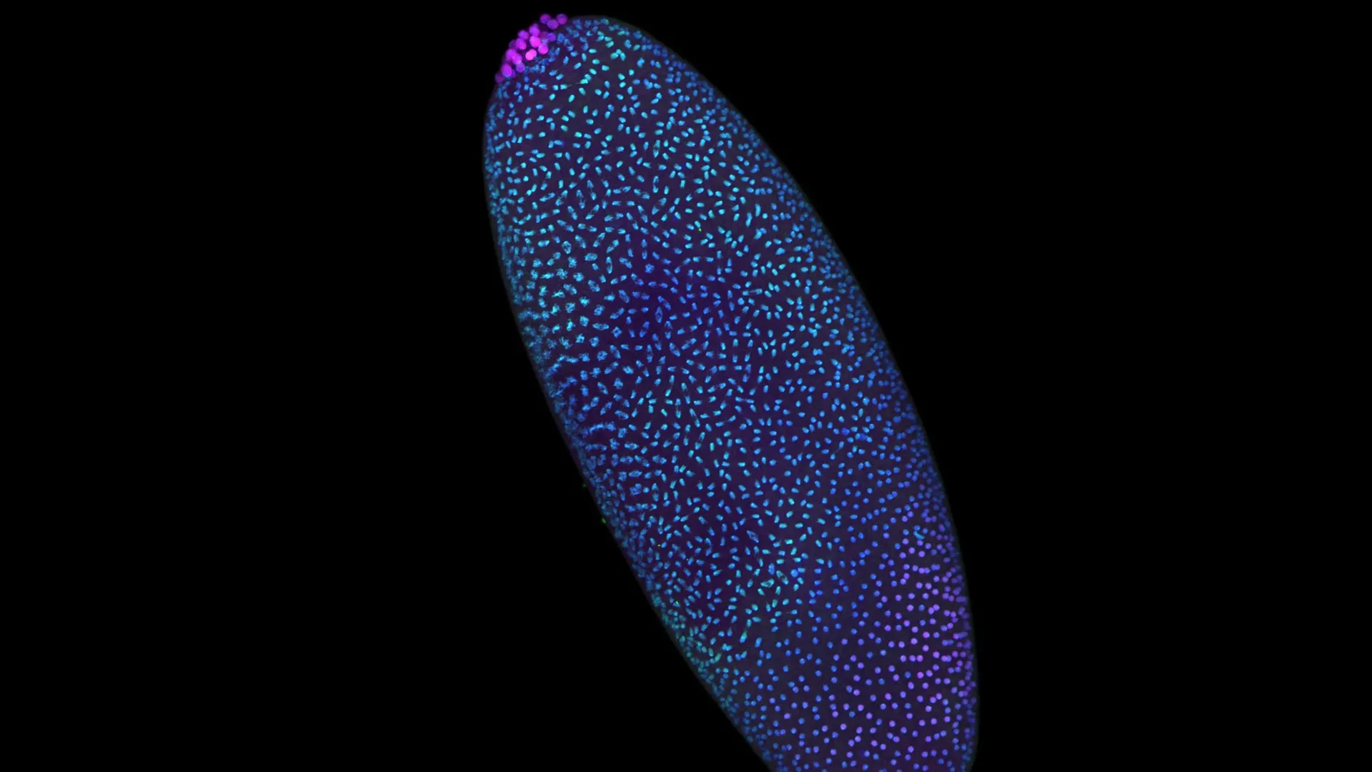
To test the capabilities of Pico-C in a living, rapidly developing system, the researchers turned to the common fruit fly (Drosophila melanogaster). The fruit fly is an iconic model organism in genetics, renowned for its fast developmental pace. During the initial hours following fertilization, a fruit fly embryo undergoes rapid, synchronous nuclear divisions, producing thousands of cells in an extraordinarily compressed timeframe.
By mapping the 3D arrangement of the fruit fly genome during these hyper-accelerated early stages using Pico-C, the research team made a startling discovery. Well before Zygotic Genome Activation occurred, the DNA was already folding into intricate, modular loops. This precise spatial arrangement dictated how regulatory signals would access specific chromosomal regions later on. Far from being a passive, tangled yarn of genetic code, the early genome was actively scaffolding itself, preparing exact architectural blueprints to ensure that genes would be switched on with absolute temporal and spatial precision.
Supporting Context, Mechanics, and Metrics
The transition from a seemingly chaotic nuclear soup to a disciplined, modular construction site relies on complex biophysical principles. To fully appreciate the significance of Professor Vaquerizas’s and Professor Kutay’s findings, one must examine the mechanics of chromatin folding and the disastrous consequences of its structural failure.
The Mechanics of Spatial Gene Regulation
DNA does not float randomly inside the cell nucleus like strands of cooked spaghetti. Instead, it is tightly packed and organized into chromatin, looping and bending to bring distant regulatory elements (such as enhancers) into physical contact with the genes they control.
- Modular Folding: The Pico-C mapping data revealed that this modular folding pattern is established proactively. The genome pre-engineers its physical layout, creating topological domains that restrict or facilitate interactions between DNA sequences.
- Precision Timing: By positioning regulatory regions near their target genes prior to activation, the cell ensures that when the transcriptional machinery finally powers up, it executes its genetic program with zero margin for error.
- Developmental Safeguards: This structural discipline is not merely a matter of efficiency; it is an absolute biological safeguard. Mislocalized or mistimed gene activation during embryogenesis frequently results in catastrophic developmental anomalies, embryonic lethality, or teratogenic malformations.
When Architecture Collapses: Insights from Human Cells
While the foundational blueprint of 3D genomics was uncovered in fruit flies, the stakes of genome architecture are equally high in human biology. In a parallel study published in Nature Cell Biology, Professor Ulrike Kutay and her collaborators at ETH Zürich in Switzerland applied high-resolution mapping strategies to human cells to observe what happens when the structural integrity of the nucleus is compromised.
The researchers targeted the molecular "anchors"—specialized proteins and nuclear envelope components—responsible for stabilizing the genome’s 3D spatial conformation. When these architectural tethers were experimentally removed, the results were both immediate and dramatic:
- Structural Catastrophe: The entire 3D framework of the genome fell apart, causing chromatin domains to unspool and lose their localized segregation.
- The Viral Mimicry Pathway: Human cells possess sophisticated innate immune surveillance mechanisms designed to detect foreign DNA (such as viral infections). When the host’s own chromatin structure collapses, stray DNA fragments and aberrant spatial configurations leak into cellular compartments where they are misidentified as viral pathogens.
- False Alarms and Chronic Inflammation: This misinterpretation tricks the cell’s innate immune system into launching an aggressive defense response. Rather than fighting off a virus, the cell triggers severe inflammatory cascades, creating a cellular environment conducive to tissue damage, degenerative disease, and oncogenesis.
[Normal Embryogenesis]
   --> Pico-C Discovery: Pre-activation 3D Scaffolding
   --> Precise Spatial Gene Regulation
   --> Normal Development & Cell Function

[Pathological Disruption]
   --> Loss of Molecular Anchors / Structural Collapse
   --> Chromatin Unspooling & Mislocalization
   --> False-Alarm Innate Immune Activation (Viral Mimicry)
   --> Chronic Inflammation & Disease
Official Statements and Expert Insights
The convergence of these two monumental studies paints a comprehensive picture of life’s fragility and organizational brilliance. The lead investigators and researchers have articulated the profound philosophical and clinical implications of their work.
Noura Maziak, lead author of the Nature Genetics study, encapsulates the shift in scientific perspective regarding the pre-activation phase:
"We used to think of the time before the genome awakens as a period of chaos. But by zooming in closer than ever before, we can see that it’s actually a highly disciplined construction site. The scaffolding of the genome is being erected in a precise, modular way, long before the ‘on’ switch is fully flipped."
Reflecting on the unified narrative told by the dual publications, Professor Juanma Vaquerizas emphasizes the tight coupling between creation and preservation:
"These two studies tell a complete story. The first shows us how the genome’s 3D structure is carefully built at the start of life. The second shows us the disastrous consequences for human health if that structure is allowed to collapse."
These expert insights underscore a unifying biological principle: life is not merely defined by the linear sequence of four nucleotide bases (A, T, C, G), but equally by the physical, three-dimensional geometry those bases occupy within the microscopic confines of the nucleus.
Future Outlook and Clinical Implications
The unveiling of pre-activation genome scaffolding and the perils of architectural collapse heralds a new era in molecular biology, genetics, and clinical medicine. As researchers harness tools like Pico-C to map chromatin dynamics with unprecedented sensitivity, several transformative frontiers are opening up.
1. Advancements in Reproductive Medicine and Embryology
Understanding that the newly fertilized egg is not a blank slate, but an intricately engineered architecture site, provides fresh diagnostic avenues for reproductive medicine. Infertility treatments, in vitro fertilization (IVF) protocols, and pre-implantation genetic diagnosis (PGD) could eventually incorporate spatial genomic evaluations. By assessing whether an early human embryo has correctly assembled its 3D chromatin scaffolding, clinicians may be able to predict developmental viability with far greater accuracy than is currently possible through morphological assessment alone.
2. Unlocking the Origins of Autoimmune and Inflammatory Diseases
The revelation from ETH Zürich that genome structural collapse triggers viral mimicry pathways opens a radical new line of inquiry into autoimmune disorders. Many chronic inflammatory conditions and laminopathies (diseases caused by mutations in nuclear envelope proteins) feature unexplained immune activation. If structural decay of the nuclear architecture routinely tricks cells into mounting immune responses against their own genetic material, therapies designed to stabilize chromatin anchors could neutralize these false alarms at their root.
3. The Future of Ultra-Low-Input Spatial Genomics
Because Pico-C requires a fraction of the biological material demanded by legacy technologies, it democratizes high-resolution structural biology. Researchers can now investigate rare cell populations—such as scarce cancer stem cells, circulating tumor cells, and minute embryonic biopsies—without losing structural resolution. This capability will accelerate discoveries in oncology, where tumor heterogeneity and structural chromosomal aberrations often drive drug resistance and metastasis.
Conclusion
The dogma of the genomic "blank slate" has officially been overturned. Through the lens of Pico-C technology, science has caught its first clear glimpse of life organizing itself from the very moment of conception. We now know that the genome builds its own house before turning on the lights, and that maintaining the structural integrity of that house is a matter of profound survival. As researchers continue to explore the breathtaking complexity of 3D nuclear architecture, humanity steps closer to mastering the foundational mechanics of life, health, and disease.