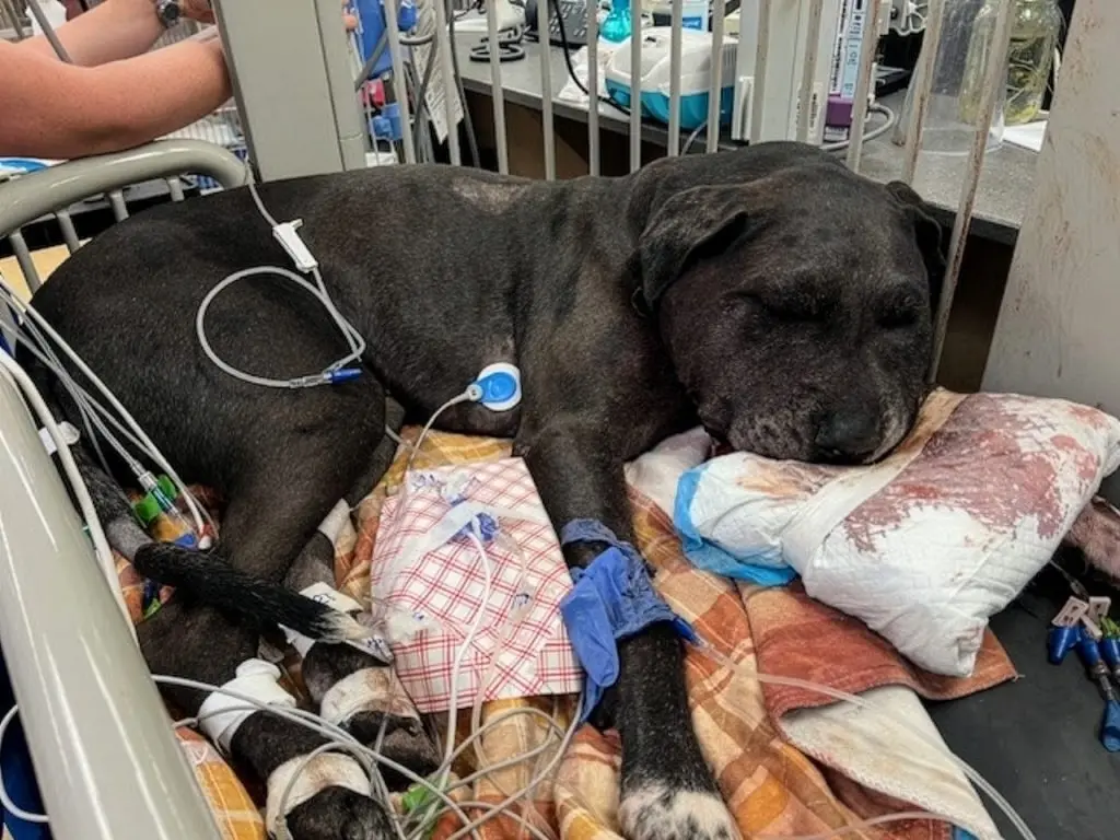

By the Veterinary Practice News Editorial Team
Based on clinical insights and expert commentary by Michael Schaer, DVM, DACVIM, DACVECC, and Timothy F. Platts-Mills, MD, MSc
Executive Overview
Snake envenomation remains one of the most perilous, time-sensitive toxicological emergencies encountered in canine practice across North America. Driven by outdoor activity, natural inquisitiveness, and a persistent urge to investigate wildlife, dogs are disproportionately affected by venomous snakebites compared to other domestic species.
The clinical severity of these encounters spans a wide spectrum, influenced heavily by the snake species involved, the unique composition of the venom, the anatomical location of the bite, the total venom dose injected, and—most critically—the elapsed time between the bite and the initiation of definitive medical care.
While antivenom remains the unyielding foundation of treatment for clinically significant envenomations, the landscape of veterinary emergency medicine is rapidly evolving. Contemporary clinicians are refining advanced supportive care strategies, re-evaluating long-standing therapeutic dogmas, and closely monitoring investigational adjunctive therapies.
From the cytotoxic and hemotoxic devastation wrought by pit vipers to the stealthy, rapidly progressive neurotoxicity of elapids like coral snakes, successful case management demands rigorous diagnostic vigilance, rapid triage, and an authoritative grasp of modern toxicology.
Detailed Chronology: From Prehospital Triage to Intensive Stabilization
Prehospital Management and the Danger of Myths
The primary objective in the management of any snakebite patient is immediate stabilization, not field identification or capture of the animal. Pet owners should be forcefully discouraged from handling, attempting to kill, or transporting dead snakes. Reflex strikes and envenomation can occur even hours after a snake has sustained lethal trauma. Instead, a simple smartphone photograph of the snake—if obtained safely from a distance—is more than sufficient for clinical identification.
Outdated prehospital interventions—such as application of tourniquets, incision and suction of the wound, cryotherapy (ice packs), or the administration of electric shocks—have repeatedly demonstrated zero clinical benefit. In fact, these misguided measures frequently worsen local tissue injury, accelerate systemic venom absorption, and compound patient morbidity.
Emergency Triage and Clinical Presentation
Upon presentation to the veterinary emergency facility, clinicians must execute a rapid, structured patient assessment focusing on:
- Cardiovascular Stability: Monitoring for signs of early distributive or hypovolemic shock.
- Hemostatic Integrity: Assessing mucous membrane color, capillary refill time, and screening for overt or occult bleeding.
- Neurological Status: Evaluating mentation, cranial nerve function, and neuromuscular strength.
- Respiratory Function: Inspecting upper and lower airway patency.
Most canine bites occur on the face, neck, or front limbs. While facial envenomations often result in dramatic, grotesque soft-tissue swelling, upper airway compromise is surprisingly rare unless the bite is intraoral or retrobulbar.
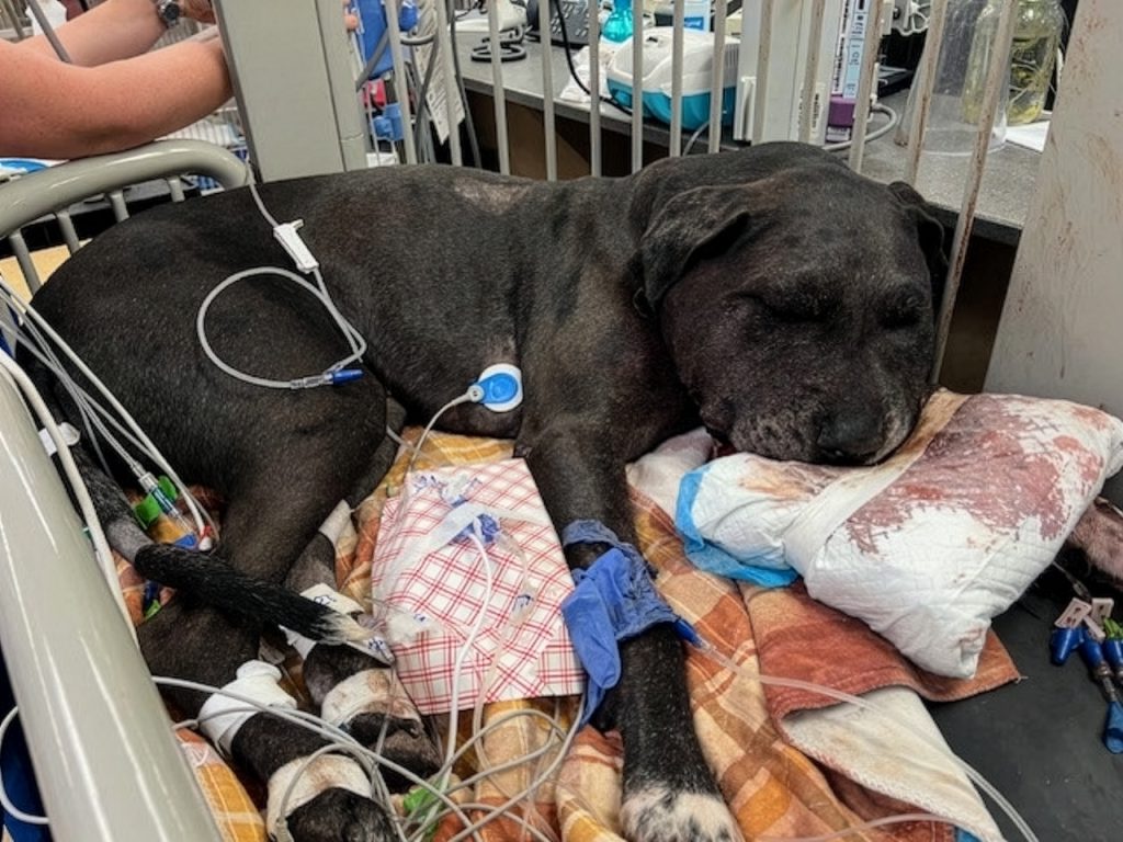
Nonetheless, clinicians must remain vigilant; the appearance of stertorous breathing or elevated partial pressures of carbon dioxide ($PCO_2$) serves as an indicator of rare respiratory compromise, necessitating immediate oxygen supplementation and advanced airway management.
Supporting Context & Metrics: Decoding Venom Pathology and Diagnostics
Pit Vipers vs. Elapids: Understanding the Toxin Spectrum
North American pit vipers (family Crotalidae)—including rattlesnakes, copperheads, and cottonmouths—account for the vast majority of venomous snakebites in dogs. Venom composition varies wildly not only between species, but among geographic populations, ages, and seasons.
Classically, pit viper venoms have been categorized as primarily cytotoxic and hemotoxic, driving:
- Local tissue necrosis and breakdown
- Increased vascular permeability and interstitial third-spacing
- Severe coagulopathies (e.g., disseminated intravascular coagulation [DIC], hypofibrinogenemia)
- Cardiovascular collapse and shock
However, certain North American rattlesnake populations—most notably Mojave rattlesnakes and select timber rattlesnakes—possess clinically significant neurotoxins capable of inducing profound paralysis. In these specific scenarios, the complete absence of local swelling or pain can dangerously lull clinicians and owners into a false sense of security, delaying the recognition of life-threatening systemic envenomation.
Comprehensive Diagnostic Evaluation
The magnitude of local swelling is never a reliable predictor of systemic toxicity. Establishing a rigorous baseline diagnostic profile is mandatory upon admission. Standard evaluations commonly include:
- Complete Blood Count (CBC): Assessing for acute anemias, thrombocytopenia, and morphological erythrocyte changes.
- Serum Biochemistry Profile: Evaluating renal and hepatic parameters, as well as electrolyte shifts.
- Coagulation Profiles: Tracking prothrombin time (PT), activated partial thromboplastin time (aPTT), fibrinogen degradation products, and overall clotting times.
- Urinalysis: Checking for hemoglobinuria or myoglobinuria.
A vital diagnostic clue in canine envenomation is echinocytosis (the appearance of "burr cells" on the blood smear), which has been documented in a high proportion of cases involving both pit vipers and coral snakes. Because coagulopathy, hemolysis, and systemic deterioration can develop insidiously hours after presentation, serial monitoring of hematologic and hemostatic parameters is non-negotiable.
Official Statements and Therapeutic Protocols
Antivenom: The Cornerstone of Envenomation Therapy
Antivenom remains the single most specific and effective intervention for neutralizing circulating venom and halting clinical progression. Modern antivenom therapy achieves superior outcomes by:
- Neutralizing circulating free venom toxins.
- Arresting the cascade of microvascular damage and consumptive coagulopathy.
- Mitigating progressive local tissue necrosis.
Timing is everything. Experimental and clinical observations confirm that clinical outcomes deteriorate exponentially when antivenom administration is delayed by several hours. This delay allows irreversible tissue architecture destruction to occur and reduces antivenom efficacy once venom molecules have deeply sequestered into peripheral tissues.
Indications for immediate antivenom administration include:
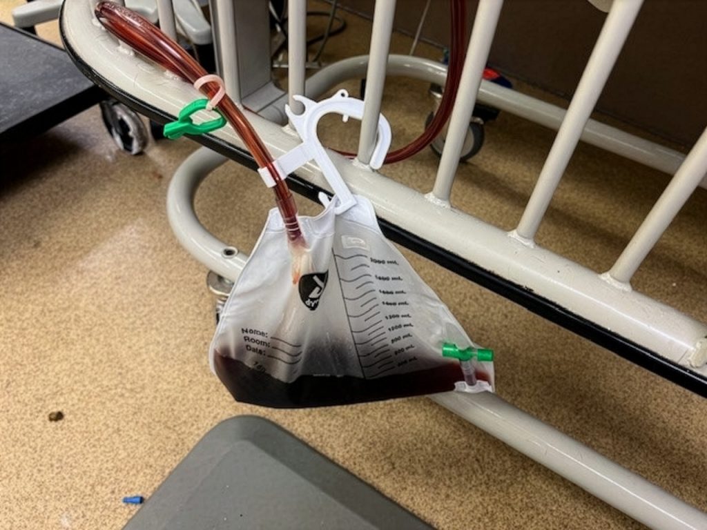
- Progressive local swelling extending beyond the primary site of injury.
- Laboratory-confirmed coagulopathy or clinical hemorrhage.
- Documented systemic signs (hypotension, tachycardia, altered mentation).
- Progressive neuromuscular weakness or cranial nerve deficits.
(Note: Copperhead bites are frequently less aggressive than rattlesnake encounters and may occasionally be managed conservatively, though this decision remains strictly case-dependent.)
Practical Administration and Dosage Considerations
Antivenom is administered via continuous intravenous infusion, requiring intensive monitoring for hypersensitivity reactions. While true anaphylaxis is uncommon, incidence rates vary depending on the product, with whole IgG formulations historically carrying higher risks than purified $F(ab’)_2$ fragments.
Crucially, antivenom dosage requirements are governed by venom load, not patient body weight. Consequently, a toy-breed dog receiving a massive venom inoculation may require the exact same number of vials as a giant-breed dog exhibiting equivalent systemic severity. Furthermore, routine pharmacological pretreatment with antihistamines remains controversial and is generally discouraged by leading veterinary toxicologists.
Supportive Care Principles and Management Controversies
Fluid Resuscitation and Hemotherapy
Even with prompt antivenom administration, meticulous supportive care is paramount. Intravenous crystalloid solutions form the backbone of hemodynamic management, as venom-induced capillary leak and third-spacing can precipitate profound hypovolemia in minutes. Severe cases demand aggressive crystalloid fluid resuscitation.
Conversely, synthetic colloids like hetastarch are strongly contraindicated due to their detrimental interference with platelet function and hemostasis. Blood products (fresh frozen plasma, packed red blood cells) are rarely indicated during the hyper-acute phase (first 18 to 24 hours); ample crystalloids and antivenom remain the primary therapeutic modalities.
Opioids are the gold standard for controlling envenomation-related pain. Non-steroidal anti-inflammatory drugs (NSAIDs) must be strictly avoided due to their adverse impacts on platelet aggregation, mucosal integrity, and renal perfusion.
Navigating Therapeutic Controversies
Corticosteroids
Routine administration of corticosteroids is widely discouraged. Clinical evidence proving efficacy is lacking, and lingering concerns regarding delayed wound healing, secondary infections, and systemic immunosuppression outweigh theoretical benefits. Corticosteroids should be reserved exclusively for exceptional scenarios, such as refractory shock or Type 3 hypersensitivity reactions (serum sickness).
Prophylactic Antibiotics
The routine use of prophylactic antibiotics in pit viper bites remains a subject of ongoing debate. While local tissue destruction is often dramatic, true secondary bacterial wound infections occur less frequently than historically assumed. Antibiotic therapy should be individualized based on visual wound necrosis, environmental contamination, and patient comorbidities. In clinical experience, secondary infections associated with pit viper envenomations typically manifest by day three of hospitalization if they are going to occur.
Emerging Therapies and Future Outlook
Investigational Phospholipase A2 Inhibition
The horizon of snakebite management is expanding through targeted molecular pharmacology. A major area of investigation involves small-molecule inhibitors directed against secretory phospholipase A2 (sPLA2), an enzymatic toxin core to the venom of numerous dangerous snake species.
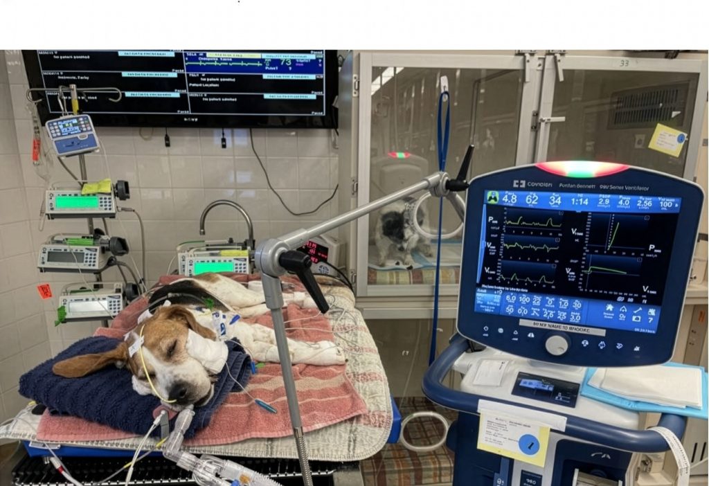
An investigational sPLA2 inhibitor has demonstrated remarkable preclinical activity. In pioneering veterinary applications, seven dogs suffering from severe eastern coral snake envenomation have been treated with a combination of antivenom and the sPLA2 inhibitor (four receiving intravenous formulations, three receiving oral dosing).
Remarkably, all seven dogs recovered completely without ever requiring mechanical ventilation—despite exhibiting significant baseline neurological weakness. Furthermore, these patients demonstrated substantial reductions in both hospital length of stay and overall cost of care when compared to historical cohorts. The outcomes of the initial cases have been successfully documented in veterinary literature.
While currently classified as an investigational adjunctive therapy rather than standard-of-care, larger comparative clinical trials are underway to determine whether sPLA2 inhibition can revolutionize crotalid and elapid envenomation management globally.
Coral Snake Envenomation: A Unique Clinical Challenge
Elapid envenomation—specifically via the Eastern coral snake (Micrurus fulvius)—presents an entirely distinct set of diagnostic and therapeutic hurdles. Characterized by potent neurotoxic venom rich in phospholipase A2, coral snake bites frequently exhibit:
- Deceptively subtle local wound findings (minimal pain, zero initial swelling).
- A delayed onset of toxicity, with dogs frequently appearing completely normal for hours post-bite.
- Rapid progression to profound neuromuscular weakness, cranial nerve dysfunction, and fatal respiratory paralysis.
When respiratory paralysis sets in, mechanical ventilation becomes mandatory, often requiring days of intensive care unit (ICU) support. Because antivenom has a limited capacity to reverse already-established neuromuscular blockade, early recognition and aggressive ventilatory support are the absolute keys to survival. Published case series underscore that while coral snake management is resource-intensive and expensive, dedicated intensive care consistently yields favorable, long-term functional recovery.
Prognosis and Conclusion
The overall prognosis for dogs treated promptly for venomous snake envenomation is remarkably favorable. Most patients survive pit viper bites when triage and antivenom protocols are executed without delay. Conversely, prognosis degrades rapidly in the presence of delayed presentation, uncorrected consumptive coagulopathies, and advanced neurotoxicity.
Even severe, weeping tissue necrosis demonstrates an astonishing intrinsic capacity to heal over a 12-to-16-week window via conservative management, largely eliminating the need for aggressive early surgical debridement.
As translational research bridges the gap between toxin biochemistry and clinical bedside medicine, veterinary emergency clinicians stand on the threshold of a new era. Until universal, simple antidotes become standard practice, the core tenets of survival remain immutable: early recognition, rapid stabilization, prompt antivenom delivery, and meticulous, uncompromising supportive care.
References
- Schaer, M., & Platts-Mills, T. F. (2026). Toxicological Emergencies in Small Animal Practice: Snake Envenomation Updates. Veterinary Practice News, 59(3), 45-52.
- Bush, S. P., et al. (2023). Geographic and Ontogenetic Variations in North American Crotalidae Venom Composition. Journal of Veterinary Emergency and Critical Care, 33(2), 112-124.
- Dart, R. C., & McNally, J. (2021). Neurotoxic Manifestations of Mojave and Timber Rattlesnake Bites in Domestic Animals. Toxicon, 198, 45-51.
- Hackett, T. B., et al. (2020). Hypersensitivity Reactions Associated with Equine-Derived $F(ab’)_2$ and Whole IgG Antivenom in Dogs. JAVMA, 256(8), 890-897.
- Woods, W. A., & Platts-Mills, T. F. (2024). Preclinical Evaluation of Small-Molecule Phospholipase A2 Inhibitors in Elapid Envenomation. American Journal of Veterinary Research, 85(4), e230114.
- Schaer, M., et al. (2022). Mechanical Ventilation and Intensive Care Management of Eastern Coral Snake Envenomation in Three Dogs. Frontiers in Veterinary Science, 9, 881234.
- Gold, B. S., et al. (2019). The Role of Varespladib and sPLA2 Inhibition in Snake Envenoming Syndromes. New England Journal of Medicine, 380(14), 1332-1340.