
Executive Overview
Deep within the structural core of the human brain rests a tiny, pinecone-shaped endocrine organ known as the pineal gland. For centuries, philosophers and scientists alike have debated its function, with René Descartes famously dubbing it the "seat of the soul." Modern neuroscience understands it primarily as the body’s internal timekeeper—a chemical factory responsible for secreting melatonin and regulating circadian rhythms in response to the cycle of day and night.
However, groundbreaking new research from an international team of biologists at Lund University and the University of Sussex suggests that the pineal gland is far more than a simple hormonal regulator. According to their findings, this obscure neural structure is the direct, living evolutionary descendant of a singular, functional sensory organ: a "median eye" that once sat squarely atop the head of our earliest vertebrate ancestors.
This revelation upends long-held orthodoxies in evolutionary biology and neuroanatomy. The study demonstrates that all vertebrates—from fish and amphibians to birds, reptiles, and mammals, including humans—can trace their lineage back to a worm-like, filter-feeding organism that roamed Earth’s primordial oceans roughly 600 million years ago. This distant relative possessed a single, central visual organ reminiscent of classical mythology’s cyclopes.
More importantly, this evolutionary detour explains a fundamental anatomical riddle that has baffled scientists for generations: why vertebrate eyes are structured so radically differently from those of invertebrates like insects and cephalopods. While a squid or a housefly develops its eyes from the ectodermal tissue of the outer skin, the vertebrate retina is born directly from the central nervous system. It is, in every sense of the word, an outpost of the brain itself.
By mapping the genetic, structural, and developmental pathways of light-sensitive cells across the animal kingdom, researchers have reconstructed a bizarre, circuitous evolutionary journey—one where our modern sense of sight was essentially rebuilt from scratch following the loss of an even older visual apparatus.
Detailed Chronology: The 600-Million-Year Evolutionary Trajectory
To understand how a sedentary, bottom-dwelling marine worm gave rise to the complex, image-forming eyes of modern vertebrates—and ultimately to the human pineal gland—we must journey back across the vast expanse of geological time to the late Precambrian era.
Phase 1: The Pre-Vertebrate Baseline and the First Reduction
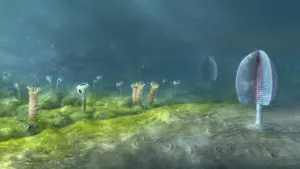
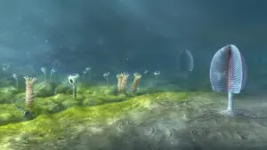
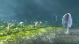
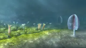
Roughly 600 million years ago, long before the explosive diversification of life known as the Cambrian explosion, the Earth’s oceans were dominated by soft-bodied invertebrate fauna. Among them was a small, elongated, worm-like organism that represented one of the earliest branches of the chordate lineage—the phylum to which all vertebrates belong.
According to evolutionary reconstructions, this primitive ancestor likely possessed a primitive set of paired eyes, much like many of its contemporary invertebrate rivals. These early visual structures may have been little more than clusters of light-sensitive photoreceptor cells or rudimentary image-forming organs capable of detecting the shifting shadows of approaching predators.
However, environmental pressures drove a radical shift in the creature’s lifestyle. Rather than actively hunting or navigating complex marine environments, the organism transitioned to a sedentary, sessile existence. It anchored itself to the ocean floor or burrowed into the sediment, surviving primarily by filtering microscopic plankton and organic detritus from passing seawater.
In this stationary ecological niche, the energetic and neurological costs of maintaining complex paired eyes outweighed their utility. Over countless generations, natural selection favored a reduction in sensory apparatus. The paired lateral eyes slowly withered and disappeared entirely.
Phase 2: The Rise of the Median Cyclopean Eye
Despite abandoning its lateral vision, the worm-like ancestor did not become entirely blind. A residual cluster of light-sensitive cells persisted in the exact center of its head, positioned dorsally (on top).
Released from the constraints of the lost paired eyes, this central cluster underwent its own evolutionary elaboration. Over millions of years, these cells organized into a simple, functional "median eye." While incapable of rendering high-resolution images of the surrounding ocean, this single median organ performed crucial survival functions: it allowed the organism to detect the broad cycle of day and night, sense the passing overhead of large shadows, and maintain spatial orientation relative to sunlight.
In essence, the earliest vertebrate relative navigated the ancient seas as a microscopic, worm-like cyclops, relying on a solitary dorsal photoreceptor to interface with the cosmos.
Phase 3: The Return to Motility and the Rebuilding of Vision
As the Earth’s geochemical and ecological landscapes shifted, paving the way for the rich evolutionary experiments of the early Paleozoic, the descendants of this sedentary organism abandoned their stationary habits. Pressured by the emergence of new, aggressive marine predators and the abundance of open-water ecological niches, these creatures returned to an active, swimming lifestyle.
This return to motility imposed severe new evolutionary demands. A swimming organism could not rely on a simple, light-detecting dorsal spot to survive; it required sophisticated spatial awareness, depth perception, and high-acuity image formation to hunt prey and evade destruction.
Faced with this biological imperative, evolution performed an extraordinary pivot. Rather than attempting to regenerate the long-lost lateral eyes of its even more distant ancestors, the organism repurposed parts of the existing median eye apparatus. Through radical genetic and developmental innovations, the region surrounding the central light-sensitive structures began to expand and differentiate, ultimately giving rise to new, paired eyes capable of advanced image formation.
Crucially, because these new visual structures budded out of the central nervous system—specifically from the neural tube that would become the brain—the resulting retinas retained their embryonic origin as brain tissue. This historical accident locked in the unique developmental trajectory of vertebrate vision for the next half-billion years.
Supporting Context & Metrics: Unraveling the Biological Blueprint
The realization that vertebrate eyes and brains share such an intertwined, unconventional history is supported by cutting-edge comparative genomics, cellular biology, and embryology.
The Retinal Anomaly: Brain Versus Skin
For decades, comparative zoologists have marveled at the stark divide between the visual systems of vertebrates and those of successful invertebrate lineages, such as arthropods (insects, crustaceans) and mollusks (octopuses, squid).
- The Invertebrate Blueprint: In insects and cephalopods, the eye develops as an invagination of the embryonic ectoderm—the outer layer of tissue that forms the animal’s skin. The photoreceptor cells and the neural tissue processing visual signals originate directly from the body surface and connect inward toward the brain.
- The Vertebrate Blueprint: In stark contrast, the vertebrate retina develops as an outward extension of the diencephalon—a region of the embryonic forebrain. The optic vesicles grow outward from the brain toward the surface of the head. Consequently, the vertebrate retina is essentially an isolated island of the central nervous system exposed to light, with neural wiring running backward relative to the direction of incoming photons.
| Feature | Vertebrate Eyes (Humans, Birds, Fish) | Invertebrate Eyes (Insects, Squid) |
|---|---|---|
| Embryonic Origin | Central Nervous System (Brain outpocketing) | Ectoderm (Surface skin tissue) |
| Retinal Wiring | Inverted (Light passes through layers of neurons) | Everted/Direct (Light hits photoreceptors directly) |
| Evolutionary Root | Rebuilt from median dorsal photoreceptors | Developed directly from surface epidermal cells |
| Ancient Remnant | Pineal Gland (Melatonin secretion, circadian regulation) | N/A (Specialized dermal/ganglionic visual organs) |
This fundamental structural discrepancy has long troubled evolutionary biologists. Standard evolutionary theory posits that complex organs evolve along linear, gradual pathways of continuous improvement. The sudden divergence in how vertebrates and invertebrates build their visual systems pointed toward a much more complicated, historically contingent past.
The new research resolves this tension. By demonstrating that vertebrate vision was effectively rebooted from a central, brain-associated median eye after the loss of ancestral lateral eyes, scientists can finally account for the "inside-out" nature of the vertebrate retina. The neural circuits responsible for analyzing images in the modern human eye trace their architectural lineage directly to the ancient neural processing centers that once interpreted signals from the 600-million-year-old cyclopean spot.
The Pineal Gland as a Living Fossil
Perhaps the most poetic confirmation of this evolutionary narrative is the persistence of the pineal gland in modern human anatomy.
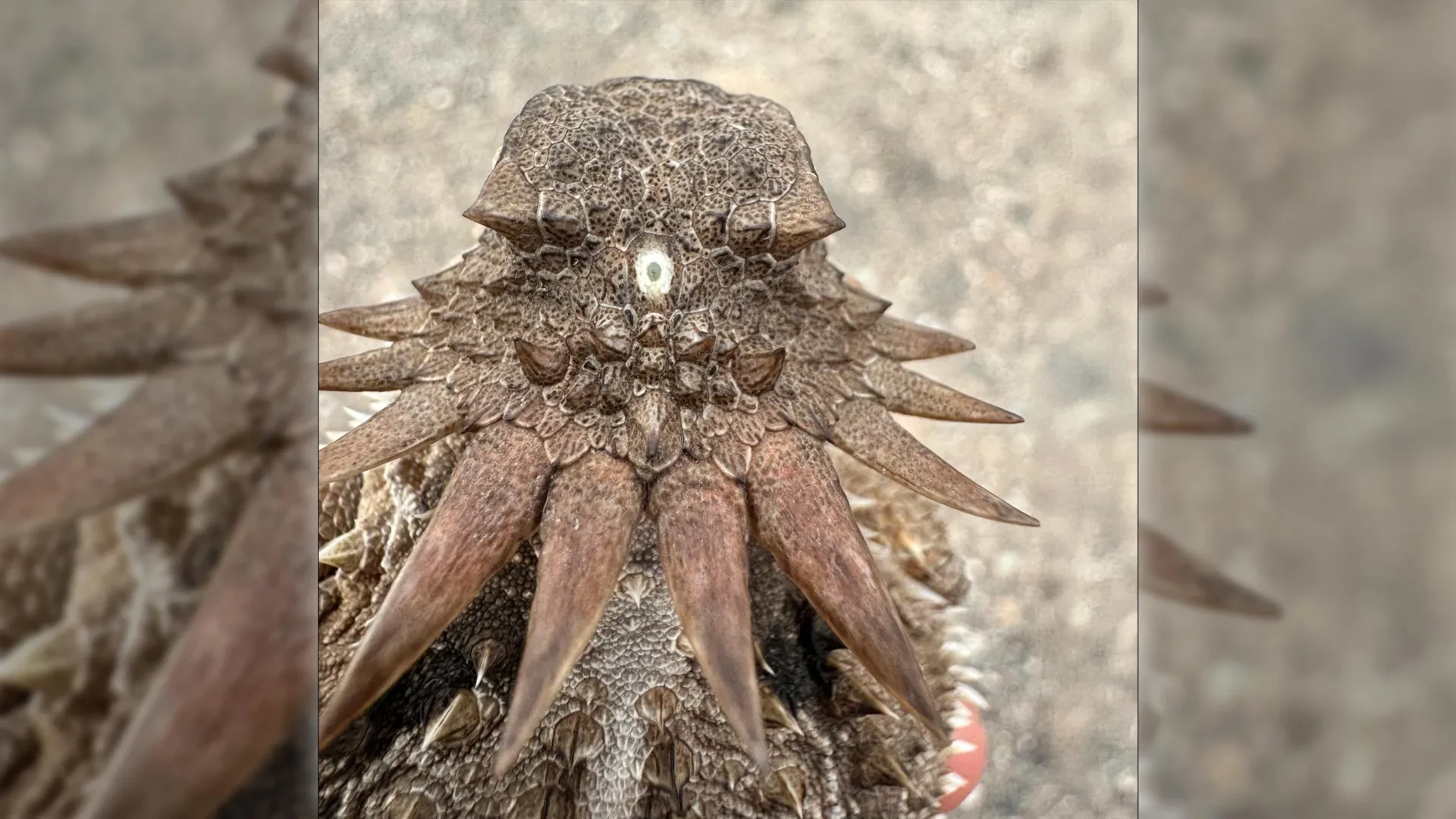
In many lower vertebrates—such as certain species of lampreys, amphibians, and reptiles—the pineal organ (sometimes referred to as the "parietal eye" or "third eye") retains a startling degree of its ancestral function. Sitting just beneath an opening in the skull, these structures possess primitive lenses, corneas, and photoreceptor cells that remain directly sensitive to sunlight, helping to regulate physiological processes without the mediation of the primary eyes.
As vertebrate evolution progressed toward mammals, the skull thickened, and the parietal eye retreated deep into the interior of the brain, losing its direct exposure to light. Yet, its cellular machinery remained intact. The pinealocytes—the primary cells of the mammalian pineal gland—evolved directly from ancient photoreceptor cells.
Even today, while human pinealocytes no longer capture photons directly through the skull, they retain the molecular machinery once used for light detection. They translate signals cascading from the modern retina via the suprachiasmatic nucleus into biochemical outputs, producing melatonin in the absence of light. Thus, every time a human being experiences the onset of sleep induced by melatonin secretion, they are utilizing a chemical signaling pathway inherited directly from a primordial, worm-like cyclops drifting through the Precambrian seas.
Official Statements and Expert Perspectives
The findings, published by researchers from Lund University and the University of Sussex, have sent ripples through the international scientific community, challenging entrenched paradigms in evolutionary developmental biology (evo-devo).
"The results are a surprise. They turn our understanding of the evolution of the eye and the brain upside down," states Dan-E Nilsson, professor emeritus in sensory biology at Lund University and one of the lead researchers on the project.
Nilsson emphasizes that the research forces a total reassessment of how complex anatomical systems can be completely rebuilt over evolutionary timescales. Rather than viewing evolution as a strictly additive process where structures are continuously refined across unbroken lineages, the study highlights the role of evolutionary loss, exaptation, and reinvention.
"Now we finally understand why the eyes of vertebrates differ so radically from the eyes of all other animal groups, such as insects and squid," Nilsson explains. "The film of our eyes—the retina—developed from the brain, whereas the eyes of insects and squid originate in the skin on the sides of the head."
The implications of the study extend far beyond structural zoology, offering profound insights into the origins of neural computation. By tracing the cellular ancestry of the vertebrate retina back to the median eye, researchers have gained unprecedented clarity on how primitive neural networks evolved to process visual data.
"For the first time, we now also understand the origin of the neural circuits that analyze the image in our retina," Nilsson adds, pointing out that the computational architecture underlying human vision was forged during the reorganization of this ancient sensory system.
Reflecting on the bizarre physiological bridge connecting modern humans to the dawn of animal life, Nilsson concludes:
"It’s mind-boggling that our pineal gland’s ability to regulate our sleep according to light stems from the cyclopean median eye of a distant ancestor 600 million years ago."
Independent evolutionary biologists have praised the rigor of the methodology, which combined high-resolution cellular mapping, comparative transcriptomics, and paleobiological reconstruction to link micro-level genetic pathways with macro-level anatomical history.
Future Outlook: What Lies Ahead for Evolutionary Neurobiology
As the dust settles on this paradigm-shifting discovery, researchers are already looking toward the horizon to explore the numerous questions the study leaves in its wake.
Mapping the Genetic Switches of Evolution
One of the primary goals for future research teams is to identify the precise genetic switches and transcription factors that governed the transition from the sessile, single-eyed phase to the active, paired-eye phase in early chordates. By utilizing advanced gene-editing techniques and single-cell RNA sequencing on living primitive vertebrates—such as amphioxus and jawless fish—scientists hope to replay the genetic tape of this ancient evolutionary transition in a laboratory setting.
Understanding how an organ as complex as the retina can be effectively re-engineered from brain-associated neural tissue could also hold transformative potential for regenerative medicine. If biomedical engineers can decode the exact molecular signals that drove the regeneration and expansion of visual structures from the central nervous system 600 million years ago, those same pathways might one day be harnessed to repair damaged human retinas or restore sight to the blind.
Expanding the Evolutionary Timeline
Furthermore, paleobiologists are intensifying their search for microfossils from the Ediacaran period that preserve soft-tissue impressions of early chordate heads. Finding physical fossil evidence of the transitional stages between paired-eye loss and median-eye dominance would provide an empirical anchor for the genomic models currently driving the field.
Ultimately, this research serves as a humbling reminder of the deep, intricate lineages that connect modern humanity to the wider tapestry of life on Earth. The eyes through which we read these words, and the internal biological clock that lulls us to sleep at night, are not isolated biological inventions. They are the living monuments of an astonishing evolutionary detour—a legacy inherited from a tiny, one-eyed wanderer of the ancient seas.