
Executive Overview
For decades, evolutionary biologists have mapped the family trees of the animal kingdom with the neat, predictable strokes of gradual progression. Yet, nature rarely takes a straight path. A groundbreaking study conducted jointly by researchers at Lund University and the University of Sussex suggests that humans, along with every other living vertebrate, are the descendants of an astonishingly bizarre creature: a tiny, worm-like marine animal featuring a single, prominent eye centered on the top of its head.
This startling hypothesis upends conventional wisdom regarding the origins of vertebrate vision. Spanning an evolutionary window nearly 600 million years ago, this "cyclopean" phase was not merely a quirky evolutionary footnote. According to the research team, this ancient, single-eyed organ directly contributed to the development of the sophisticated paired eyes we use today. Even more remarkably, evolutionary remnants of this ancient visual system appear to persist within the modern human brain as the pineal gland—a vital endocrine structure that regulates our sleep-wake cycles.
"The results are a surprise. They turn our understanding of the evolution of the eye and the brain upside down," says Dan-E Nilsson, professor emeritus in sensory biology at Lund University and a leading voice behind the study.
By combining broad comparative analyses of light-detecting cells, neural circuitry, and developmental biology, the international research team has pieced together an intricate evolutionary puzzle. Their findings explain long-standing anatomical mysteries, including why vertebrate eyes are structurally inverted compared to those of invertebrates like insects and cephalopods, and how our neural circuits for processing light first came into being. This report explores the chronological sequence of this evolutionary detour, examines the supporting scientific context, analyzes expert commentary, and evaluates the broader implications for evolutionary science.
Detailed Chronology: The 600-Million-Year Evolutionary Trajectory
To understand how humans evolved from a one-eyed marine organism, scientists must peer deep into the Ediacaran and early Cambrian periods, an era when complex multicellular life was rapidly diversifying in Earth’s primordial oceans.
Phase I: The Shift to a Sedentary Existence
Nearly 600 million years ago, the distant ancestor of all vertebrates was a modest, soft-bodied creature bearing a morphological resemblance to a modern marine worm. This organism inhabited shallow benthic environments, anchoring itself or moving sluggishly along the ocean floor. Its primary mode of survival was filter feeding, extracting suspended plankton and organic detritus from the surrounding seawater.
Earlier in its lineage, this organism—or its immediate forebears—likely possessed paired eyes or bilateral clusters of light-sensitive cells. In actively moving animals, paired eyes confer immense evolutionary advantages: they allow for stereoscopic depth perception, the precise judgment of distance, and the rapid tracking of moving predators or prey across a dynamic visual field.
However, as the creature adopted a sedentary, filter-feeding lifestyle, the survival pressures driving the maintenance of complex paired vision evaporated. Navigating a complex spatial environment was no longer a daily priority. Over countless generations, natural selection relaxed its grip on these lateral visual structures. Lacking any significant functional advantage, the paired eyes gradually regudated and disappeared from the organism’s morphology.
Phase II: The Rise of the Median Eye
While the lateral visual structures faded away, the organism retained a localized cluster of photoreceptor cells positioned squarely in the middle of its head. This cluster evolved into a functional median eye—a primitive, single-eyed structure reminiscent of mythological cyclopeans.
Because this median eye lacked a complex lens and high-resolution focusing mechanics, it could not form detailed, high-definition images of the surrounding environment. Instead, it served a more fundamental ecological purpose: detecting ambient light levels, distinguishing between day and night, and sensing the vertical axis of the water column (determining which direction was upward toward the safety of the sunlit surface). For a stationary filter feeder, this basic photosensitivity was more than sufficient to coordinate daily biological rhythms and avoid surface hazards.
Phase III: The Return to Mobility and the Invention of Vertebrate Eyes
Millions of years passed, and environmental pressures shifted once more. The descendants of this sedentary worm-like ancestor abandoned their stationary habits, returning to an active, swimming lifestyle.
A mobile existence immediately resurrected the need for sophisticated vision. Navigating open waters required the rapid detection of food sources, the identification of physical obstacles, the evasion of agile predators, and the ability to track directional movement. Rather than reinventing vision from scratch, evolution performed an extraordinary morphological recycling project.
The researchers conclude that portions of the original median eye were co-opted and repurposed. Through a complex series of developmental shifts, this central visual structure acted as the developmental template from which a new pair of image-forming eyes emerged. This unusual evolutionary detour explains a fundamental anatomical riddle that has vexed biologists for generations: why vertebrate eyes are structurally inverted compared to the eyes of insects, crustaceans, and cephalopods like squid.
In insects and squid, the retina—the light-sensitive tissue lining the eye—develops outward from the ectodermal skin tissue on the sides of the head. In stark contrast, the vertebrate retina develops directly from neural tissue within the embryonic brain. Because our evolutionary lineage passed through a stage where the visual apparatus was centralized on the head’s dorsal surface, the retina originated as an outgrowth of the central nervous system. This deep neuro-developmental lineage also explains why vertebrate retinas contain intricate internal neural circuits that begin processing visual data before the signals are even transmitted to the higher centers of the brain.
Supporting Context & Metrics: Decoding the Evidence
The conclusions drawn by the Lund University and University of Sussex teams are not based on a single fossil discovery; rather, they stem from a massive, multi-disciplinary synthesis of comparative biology, neuroanatomy, and embryology.
Comparative Photoreceptor Mapping
To reconstruct this evolutionary path, the researchers conducted an exhaustive comparative analysis of light-detecting cells across nearly all major animal phyla. By mapping where these cells appear anatomically, analyzing their biochemical pathways, and tracing how they wire into adjacent nervous systems, the team identified distinct evolutionary lineages of vision.
The data revealed that while arthropod and mollusk eyes evolved via epidermal invagination (skin folding inward), the vertebrate visual system bears the unmistakable biochemical and neurological signatures of brain-derived photoreception. This clear demarcation supports the hypothesis of an independent, highly specialized evolutionary origin for vertebrates.
The Survival of the Median Eye: The Pineal Gland
Perhaps the most astonishing component of this research is the identification of the ancient median eye’s modern descendant. The single eye did not vanish entirely; instead, it retreated deep into the interior of the cranium.
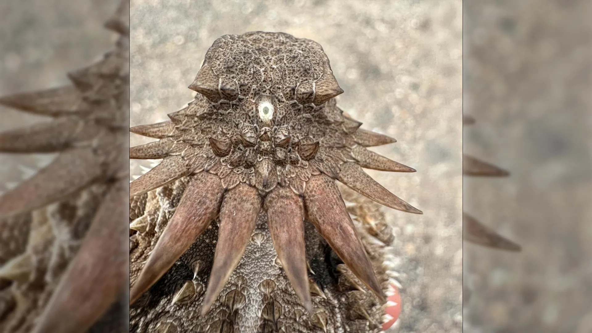
In modern vertebrates, including humans, this evolutionary relic survives as the pineal gland—a tiny, pinecone-shaped endocrine organ nestled between the two hemispheres of the brain. While human pineal glands no longer form images or directly register bright light through the skull, they retain deep biochemical ties to our ancient photosensory heritage.
| Feature / Organ | Ancient Median Eye | Modern Human Pineal Gland |
|---|---|---|
| Anatomical Position | Central, dorsal surface of the head | Deep interior of the brain (epithalamus) |
| Primary Function (Ancestral) | Detecting day/night cycles, vertical orientation | Regulating endocrine rhythms and sleep architecture |
| Photosensitivity | Direct light detection via primitive photoreceptors | Indirectly regulated by light signals routed from the retinal eyes |
| Hormonal Output | Unknown / Primitive signaling molecules | Synthesis and secretion of melatonin |
In humans and many other mammals, light information is captured by the paired eyes, converted into neural impulses, and transmitted via the retinohypothalamic tract to the suprachiasmatic nucleus, which in turn commands the pineal gland. When ambient light fades, the pineal gland ramps up production of melatonin—the key hormone responsible for inducing drowsiness and maintaining the circadian rhythm across a 24-hour cycle. In many lower vertebrates (such as certain fish, amphibians, and reptiles), the pineal organ retains a direct, skull-penetrating sensitivity to light, acting virtually as a "third eye."
Official Statements and Expert Perspectives
The implications of this study stretch far across the landscape of evolutionary biology, challenging long-held assumptions regarding how complex anatomical systems are assembled and modified over deep geological time.
Professor Dan-E Nilsson, lead researcher and professor emeritus in sensory biology at Lund University, emphasizes the paradigm-shifting nature of the discovery:
"The results are a surprise. They turn our understanding of the evolution of the eye and the brain upside down. For the first time, we now also understand the origin of the neural circuits that analyze the image in our retina."
Nilsson underscores the profound cognitive dissonance of realizing that a fundamental human biological mechanism—our sleep regulation—is directly linked to an archaic, single-eyed marine organism:
"It’s mind-boggling that our pineal gland’s ability to regulate our sleep according to light stems from the cyclopean median eye of a distant ancestor 600 million years ago."
Other members of the research consortium highlight how this model resolves persistent debates regarding vertebrate eye architecture. By demonstrating that the retina is fundamentally an extension of the brain rather than specialized skin tissue, the study provides a unified framework for understanding why vertebrate eyes function with a level of internal signal preprocessing that distinguishes them sharply from invertebrate phyla.
Future Outlook: Implications for Evolutionary Biology and Medicine
As the scientific community digests these findings, the research opens several critical avenues for future investigation:
- Genomic and Developmental Mapping: Researchers aim to trace the specific genetic toolkits (such as Pax6 and related master control genes for eye development) across basal chordates like lancelets and tunicates to pinpoint the exact molecular triggers that governed the transition from a median eye to paired lateral structures.
- Reconstructing Ediacaran Ecology: Paleobiologists are re-evaluating Ediacaran fossil beds with renewed focus on micro-anatomy, searching for soft-tissue impressions that might preserve transitional stages of early chordate neuro-anatomy.
- Chronobiological and Medical Insights: Understanding the deep evolutionary roots of the pineal gland and melatonin synthesis may offer fresh perspectives on modern circadian disorders, jet lag, seasonal affective disorder (SAD), and the neurochemical mechanisms governing human sleep architecture.
Ultimately, this discovery serves as a humbling reminder of nature’s evolutionary creativity. The human eye—an organ celebrated by poets and scientists alike as a pinnacle of biological engineering—traces its lineage back through a bizarre, winding path: from active swimmers to blind, sedentary filter-feeders, through a bizarre single-eyed cyclopean phase in the murky oceans of the Precambrian, and finally into the intricate neural architecture that allows us to read these words today.