
Executive Overview
For decades, evolutionary biologists have mapped the family tree of vertebrates—a vast lineage encompassing humans, birds, reptiles, amphibians, and fish—using genetic sequencing, fossil records, and comparative anatomy. Yet, foundational questions concerning the origin of our sensory organs have remained stubbornly unresolved. Chief among these is the stark anatomical divergence between the sophisticated, brain-derived eyes of vertebrates and the surface-derived ocular systems of invertebrates like insects and cephalopods.
Groundbreaking research from Lund University and the University of Sussex proposes a radical paradigm shift. According to the study, the earliest ancestors of all living vertebrates passed through a bizarre, "cyclops-like" phase approximately 600 million years ago. During this epoch, a tiny, worm-like marine organism abandoned its active lifestyle for a stationary existence, systematically discarding its paired eyes. In their place, it retained a solitary, centrally located median eye atop its head.
Millennia later, as descendants of this creature returned to an active, swimming lifestyle, evolutionary pressures demanded a renewed visual apparatus. Remarkably, rather than regenerating lost systems from scratch, evolution repurposed this central median structure to forge an entirely new set of paired eyes. This extraordinary detour explains why the vertebrate retina develops directly from brain tissue—a distinct developmental trajectory not shared by insects or squid.
Even more astonishingly, the physical remnants of this ancient median eye have not vanished from the biological ledger. Instead, they survive deep within the modern human brain as the pineal gland, an endocrine organ responsible for regulating our sleep-wake cycles and circadian rhythms in response to light. This synthesis of paleontology, developmental biology, and comparative neuroanatomy upends long-held orthodoxies regarding how human sight and neurological systems evolved.
Detailed Chronology: The 600-Million-Year Visual Odyssey
To trace the lineage of vertebrate vision, scientists must peer deep into the Ediacaran period, a critical juncture in Earth’s history when complex multicellular life began to proliferate in the world’s oceans. The events that sculpted the modern human eye unfolded across several major evolutionary phases.
Phase I: The Primal Marine Ancestor and the Loss of Paired Sight
Nearly 600 million years ago, the proto-vertebrate lived on the ocean floor. Possessing a soft, worm-like body, this primitive creature fed by filtering passing plankton from seawater, adopting a largely sedentary, benthic lifestyle.
Paleontological and comparative genetic data suggest that this organism’s even earlier forebears possessed paired light-sensitive cell clusters or rudimentary eyes. In mobile organisms, paired eyes confer distinct evolutionary advantages by facilitating directional judgment, spatial awareness, and distance calculation. However, for a stationary filter-feeder, maintaining complex paired eyes proved energetically costly and functionally redundant.
As generations passed, natural selection relaxed its pressure on the paired visual structures. Without a behavioral need to hunt, evade fast-moving predators, or navigate complex topographies, the organism gradually lost its lateral eyes. The metabolic investment required to maintain paired visual pathways outweighed any survival benefits, leading to their evolutionary regression.
Phase II: The Cyclopean Bottleneck
While the lateral eyes faded into history, the ancient organism retained a distinct cluster of light-sensitive cells positioned centrally on top of its head. This yielded a primitive median eye—often characterized by researchers as a "cyclopean" stage.
Though incapable of resolving sharp images or processing high-resolution visual scenes, this median eye served critical survival functions. It provided rudimentary phototaxis (movement toward or away from light), enabled the organism to distinguish between day and night, and supplied a reliable sense of vertical orientation (knowing which direction led up toward the surface and safety).
This central light-sensing organ acted as a vital evolutionary holding pattern. For millions of years, the lineage sustained itself through this simplified sensory mechanism, anchoring the structural blueprint for future visual innovations.
Phase III: The Return to Mobility and the Genesis of Paired Retinas
As global ecosystems shifted and ecological niches expanded, descendants of this sedentary worm-like creature re-embarked on an active, swimming lifestyle. A mobile existence introduced complex behavioral demands: hunting prey, avoiding dynamic hazards, and processing rapid spatial changes.
Faced with a sudden demand for advanced vision, evolution executed an unprecedented architectural maneuver. Rather than reverting to the ancient invertebrate design of forming eyes from the outer epidermal tissue (as seen in insects and mollusks), the organism repurposed portions of its existing central median eye structure.
Through complex genetic and embryological shifts, the central light-detecting tissue expanded and re-differentiated, giving rise to a new pair of image-forming eyes. Because this developmental cascade originated from cells already integrated with the central nervous system, the resulting light-sensitive membrane—the retina—formed as an extension of the embryonic brain itself. This embryological origin explains why the vertebrate retina remains structurally continuous with the central nervous system throughout life.
Supporting Context & Metrics: Comparative Anatomy and Neural Architecture
The implications of this discovery extend far beyond paleontological curiosity, shedding light on fundamental differences in neurobiology across the animal kingdom.
The Developmental Divide: Vertebrates vs. Invertebrates
To understand the significance of the Lund-Sussex findings, one must examine the embryogenesis of ocular structures across disparate phyla:
- Invertebrates (e.g., Insects, Cephalopods): The eyes of animals like Drosophila (fruit flies) and squids originate from the ectoderm—the outer layer of embryonic tissue that forms the skin. Ocular development is driven by surface invaginations and epidermal differentiation, connecting to the brain via secondary nerve tracts.
- Vertebrates (e.g., Humans, Mammals, Fish): The vertebrate eye develops via an outgrowth of the diencephalon (part of the embryonic forebrain). The optic vesicle pushes outward toward the surface ectoderm, folding inward to form the multilayered neural retina.
This deep developmental divergence has puzzled biologists for decades. The hypothesis that vertebrate eyes emerged from a repurposed median central eye neatly resolves this riddle. It demonstrates that vertebrate vision is not a modification of the standard invertebrate optic plan, but the product of an internal neural structure that migrated, duplicated, and specialized.
Neural Circuitry and Information Processing
The study also illuminates the origins of retinal neural circuits. In modern vertebrates, the retina does not merely act as a passive camera film; it is an active computational center. Specialized networks of bipolar cells, horizontal cells, amacrine cells, and retinal ganglion cells process contrast, motion, and spatial frequency before visual signals ever reach the visual cortex of the brain.
By establishing that the vertebrate retina evolved from pre-existing central brain structures, researchers can now trace the stepwise assembly of these sophisticated neural circuits. The genetic toolkits responsible for sorting visual information were already operating within the primitive light-sensing apparatus of our 600-million-year-old ancestors, providing a pre-adapted foundation for advanced image processing.
Official Statements and Expert Analysis
The paradigm-shifting nature of the research has drawn widespread attention within the global scientific community. Professor Dan-E Nilsson, professor emeritus in sensory biology at Lund University and a leading author of the study, emphasized the disruptive nature of their findings during a recent presentation of the work.
"The results are a surprise. They turn our understanding of the evolution of the eye and the brain upside down," Nilsson stated.
Reflecting on the distinct structural dichotomy separating human vision from that of other complex organisms, Nilsson elaborated on the mechanics of the vertebrate retina:
"Now we finally understand why the eyes of vertebrates differ so radically from the eyes of all other animal groups, such as insects and squid. The film of our eyes—the retina—developed from the brain, whereas the eyes of insects and squid originate in the skin on the sides of the head."
Addressing the astonishing neurological persistence of our ancient ancestry, Nilsson highlighted the connection between primordial light detection and modern human physiology:
"For the first time, we now also understand the origin of the neural circuits that analyze the image in our retina… It’s mind-boggling that our pineal gland’s ability to regulate our sleep according to light stems from the cyclopean median eye of a distant ancestor 600 million years ago."
Co-researchers from the University of Sussex added that comparative genomic mapping and modern embryological tracing techniques were instrumental in validating the model. By cross-referencing light-sensing opsin proteins across diverse marine phyla, the research team successfully mapped the genetic continuity linking Ediacaran filter-feeders to modern chordates.
The Ancient Eye Inside Modern Humans: The Pineal Gland
Perhaps the most profound takeaway for the general public is the realization that a piece of this ancient cyclopean eye remains active inside the human skull today.
Deep within the epithalamus, nestled between the two hemispheres of the brain, lies the pineal gland—a small, pinecone-shaped endocrine structure. While human beings do not consciously "see" images through the pineal gland, its physiological function remains intimately tied to light perception.
Melatonin and Circadian Regulation
The pineal gland’s primary function is the synthesis and secretion of melatonin, a serotonin-derived hormone that modulates sleep patterns in both circadian and seasonal cycles.
- Darkness Response: When environmental light fades, specialized intrinsically photosensitive retinal ganglion cells (ipRGCs) in the human eye signal the suprachiasmatic nucleus (SCN) of the hypothalamus. The SCN relays this information to the pineal gland, triggering an up-regulation in melatonin production. This hormonal surge induces drowsiness, lowering core body temperature and preparing the organism for sleep.
- Light Suppression: Conversely, exposure to morning light halts melatonin synthesis, promoting alertness and synchronizing the body’s internal biological clock to the external solar day.
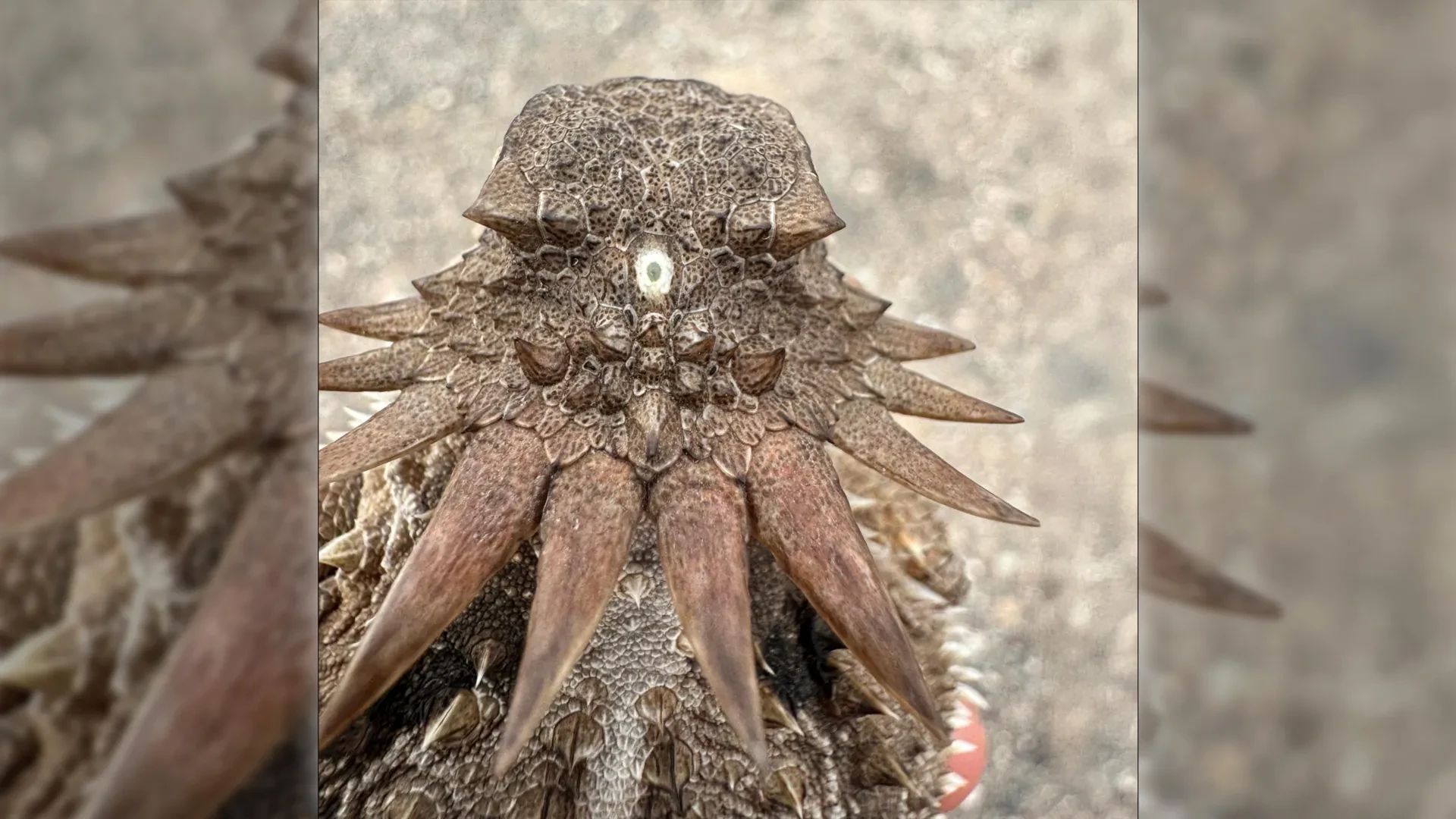
In lower vertebrates—such as certain species of fish, amphibians, and reptiles—the pineal organ (often referred to as the "parietal eye" or "third eye") retains direct, unmediated photosensitivity, lying just beneath a translucent patch of skull. While evolutionary pressures in mammals drove this structure inward and insulated it from direct ambient light, the underlying biochemical machinery persisted.
Thus, every time a human experiences jet lag, grapples with seasonal affective disorder (SAD), or simply feels tired at bedtime, they are engaging a neurochemical pathway inherited directly from a one-eyed marine organism that drifted through prehistoric oceans 600 million years ago.
Future Outlook: Unresolved Questions and Next Steps
While the Lund-Sussex study provides a compelling framework for vertebrate eye evolution, it also opens new avenues for ongoing investigation within evolutionary developmental biology ("evo-devo") and neurobiology.
Genomic and Fossil Exploration
Researchers are currently expanding their comparative genetic analyses to identify the precise regulatory genes that controlled the transition from the median eye to paired retinal outgrowths. By sequencing the genomes of basal chordates—such as lancelets (Amphioxus) and tunicates—scientists hope to isolate transitional genetic switches that governed this anatomical remodeling.
Furthermore, paleontologists are re-examining Ediacaran and early Cambrian fossil beds for micro-fossils bearing preserved soft-tissue impressions of proto-vertebrate heads. Finding physical evidence of intermediate stages between the cyclopean bottleneck and the re-emergence of paired eyes would provide definitive empirical validation for the model.
Clinical and Neurological Implications
Understanding the shared evolutionary origin of the retina and the brain may also yield long-term benefits for regenerative medicine. By mapping how neural tissue successfully invaginated and differentiated into complex visual circuits during deep evolutionary time, biomedical engineers hope to gain critical insights for repairing damaged human retinas, treating degenerative neural conditions, and engineering bio-synthetic optical interfaces.
As science continues to peel back the layers of our biological history, the boundary between myth and evolutionary reality blurs. The ancient cyclops of ancient seas is not merely a figment of evolutionary folklore; it is the direct structural architect of human sight and the silent guardian of human sleep.