
Executive Overview
Deep within the evolutionary lineage shared by humans and every other living vertebrate lies a startling and counterintuitive origin story: a microscopic, worm-like marine organism that lived nearly 600 million years ago, featuring a single, central eye mounted squarely on top of its head.
Groundbreaking collaborative research conducted by evolutionary biologists at Lund University and the University of Sussex has turned decades of established embryological and anatomical dogma on its head. According to their findings, the earliest ancestors of the vertebrate subphylum passed through a distinct, cyclops-like evolutionary bottleneck. During this phase, the organism discarded its primitive paired eyes—organs rendered redundant by a sedentary, filter-feeding lifestyle—and relied exclusively on a solitary median eye to navigate its primordial environment.
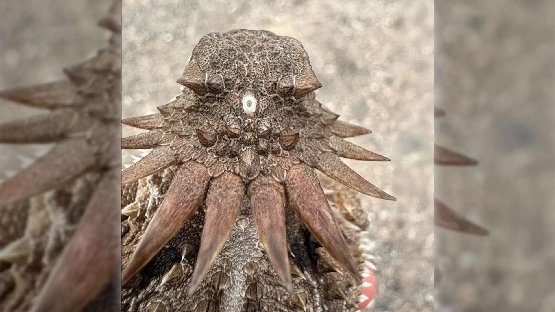
Even more remarkably, the scientific team posits that this ancient visual system did not vanish into the evolutionary ether. Instead, physical remnants of this median eye appear to persist deep within the modern human brain as the pineal gland—a neuroendocrine organ chiefly responsible for regulating circadian rhythms and sleep-wake cycles through the production of melatonin. Furthermore, this bizarre evolutionary detour explains one of comparative zoology’s most perplexing anomalies: why vertebrate eyes, featuring retinas that develop directly out of brain tissue, are constructed in a manner fundamentally opposed to the surface-tissue eyes found in invertebrates such as insects and cephalopods.
This report explores the mechanisms of this extraordinary evolutionary journey, synthesizing the morphological evidence, official academic statements, and broader biological implications of a discovery that bridges the gap between deep-sea prehistory and contemporary neuroscience.
Detailed Chronology: The Three-Act Evolutionary Saga of the Vertebrate Eye
To comprehend how humanity and its distant vertebrate cousins inherited an optical architecture rooted in a cyclopean blueprint, researchers trace a three-act evolutionary drama spanning hundreds of millions of years. This timeline details how environmental pressures shaped, dismantled, and ultimately reinvented the biological apparatus used to perceive the world.
Act I: The Loss of the Paired Vision (Pre-600 Million Years Ago)
Long before the advent of jawed fish, tetrapods, mammals, and primates, the shared stem-ancestor of all vertebrates was a small, soft-bodied marine invertebrate. Early in this organism’s ancestral lineage, genetic blueprints successfully coded for paired, lateral light-sensitive structures. In the animal kingdom, paired eyes are considered an evolutionary gold standard. By positioning light-detecting cell clusters on opposite sides of a mobile body, an organism can efficiently calculate direction, gauge physical distance, and track the movement of predators or prey.
However, environmental adaptations often supersede ancestral advantages. Over successive generations, this specific ancient lineage adopted a remarkably sedentary, benthic lifestyle. Rather than actively hunting or navigating complex oceanic terrain, the organism anchored itself to the seafloor, existing primarily as a filter-feeder. It strained microscopic plankton and organic particulate matter directly from passing ocean currents.
Because the animal spent the vast majority of its existence stationary, the immense metabolic and genetic cost of maintaining complex, paired lateral eyes offered diminishing returns. Without the selective pressure of motion-based survival, the genetic pathways governing the development of these lateral structures withered. Across deep time, the paired eyes completely disappeared from the organism’s anatomical profile.
Act II: The Reign of the Median Eye (Approximately 600 Million Years Ago)
While the lateral eyes regressed, a separate cluster of photoreceptive cells situated squarely in the middle of the animal’s head remained intact. Free from the biological real estate occupied by the lost paired organs, this central cluster expanded and developed into a functional, primitive median eye—effectively transforming the creature into a biological cyclops.
Though incapable of forming high-resolution, detailed images or discerning the fine features of objects, this median organ served vital ecological functions. It provided the stationary organism with crucial environmental awareness:
- Circadian Orientation: The cell cluster allowed the creature to reliably distinguish between day and night, synchronizing its metabolic processes with the rotation of the Earth.
- Geotactic Navigation: By detecting the direction from which ambient sunlight filtered down through the water column, the median eye provided a dependable vertical reference, enabling the organism to determine which direction was upward.
This singular, central visual system sustained the lineage through a critical evolutionary bottleneck, proving more than adequate for a quiet, stationary existence on the Precambrian seafloor.
Act III: The Great Re-Emergence and Retinal Reconstruction
Millions of years later, environmental shifts and ecological opportunities prompted descendants of this sedentary organism to abandon their stationary habits. A new generation of animals took to active swimming, hunting, and exploring the dynamic waters of the ancient ocean.
This return to a mobile lifestyle created an immediate, high-stakes evolutionary demand for advanced vision. The animal once again needed to detect obstacles, spot approaching predators, locate dispersed food sources, and navigate fluid trajectories in three-dimensional space.
Rather than restarting ocular evolution from scratch, nature resorted to genetic recycling. The researchers conclude that the organism co-opted portions of its existing median eye structure, repurposing its neural architecture to construct an entirely new pair of image-forming eyes. This unusual recycling project accounts for the distinct embryological origin of vertebrate vision. While insects, spiders, and marine squids evolved their eyes from the ectodermal tissue on the exterior surface of the head, vertebrate retinas developed directly out of evaginations of the embryonic brain itself. The retina—the intricate layer of neural tissue lining the interior back of the vertebrate eye—is quite literally a piece of the central nervous system pushed outward into the light.
Supporting Context & Metrics
The validity of this evolutionary model rests on an exhaustive, comparative analysis of light-detecting cells, neural wiring diagrams, and anatomical structures across diverse biological phyla.
Comparative Anatomy: Vertebrates vs. Invertebrates
To appreciate the significance of the Lund-Sussex findings, scientists look at the structural divergence between vertebrate and invertebrate optical systems:
| Metric / Feature | Vertebrate Eyes (Humans, Fish, Birds) | Invertebrate Eyes (Insects, Cephalopods) |
|---|---|---|
| Embryological Origin | Outgrowth of the developing embryonic brain | Infolding of surface ectoderm (skin tissue) |
| Retinal Structure | "Inverted" (light must pass through layers of nerve cells before hitting photoreceptors) | "Eververted" (photoreceptors face the light source directly) |
| Neural Processing | Initial visual signal processing occurs locally within retinal neural circuits before reaching the brain | Processing largely deferred until signals arrive at the central optic lobes |
| Evolutionary Root | Repurposed median-eye structures from a cyclopean ancestor | Independent parallel evolution from surface light-spots |
The Mechanics of Retinal Neural Circuits
One of the most profound revelations of the new study involves the origin of retinal neural networks. In standard evolutionary theory, the complex web of neurons that pre-processes visual data—sorting edges, calculating contrast, and tracking motion before signals ever travel down the optic nerve—was thought to have assembled spontaneously alongside the vertebrate eye.
By tracing the lineage back to the median eye of the cyclopean ancestor, researchers now understand that these intricate neural circuits predate the modern paired eyes. They evolved first to process simple light signals in the central organ and were subsequently duplicated and expanded when the new paired, image-forming eyes emerged. This explains why human vision possesses built-in computational power right at the back of the eyeball: the machinery was inherited from an ancient processing center that had already spent millions of years optimizing light data.
Official Statements & Academic Perspectives
The implications of this study have sent ripples through the international evolutionary biology community. By upending long-held assumptions about how the brain and eye co-evolved, the research demands a revision of standard academic textbooks.
Dr. Dan-E Nilsson, professor emeritus in sensory biology at Lund University and a leading voice behind the research, expressed astonishment at where the data led his team:
"The results are a surprise. They turn our understanding of the evolution of the eye and the brain upside down," Professor Nilsson stated, emphasizing the radical departure from traditional phylogenetic models.
Reflecting on the persistence of the ancient median eye within modern anatomy, Nilsson highlighted the sheer improbability of human neurobiology:
"It’s mind-boggling that our pineal gland’s ability to regulate our sleep according to light stems from the cyclopean median eye of a distant ancestor 600 million years ago."
Furthermore, Nilsson underscored how this discovery solves a decades-old comparative anatomy puzzle that has vexed generations of zoologists:
"For the first time, we now also understand the origin of the neural circuits that analyze the image in our retina… Now we finally understand why the eyes of vertebrates differ so radically from the eyes of all other animal groups, such as insects and squid. The film of our eyes—the retina—developed from the brain, whereas the eyes of insects and squid originate in the skin on the sides of the head."
Independent evolutionary developmental biologists have praised the study for synthesizing molecular genetics, comparative neuroanatomy, and fossil-record inferences into a cohesive, testable hypothesis. While controversies inevitably arise when shifting foundational paradigms, the clarity provided regarding the pineal gland and retinal ontogeny has earned widespread professional respect.
Future Outlook & Broader Implications
As the scientific community digests the conclusions put forth by the Lund University and University of Sussex research teams, the focus is shifting toward future avenues of investigation.
Genomic Archaeology and Molecular Tracers
Researchers are currently planning molecular mapping studies to search for conserved genetic toolkits across modern chordates and basal invertebrates (such as lancelets and tunicates). By examining the exact regulatory genes that govern both pineal gland development and retinal differentiation, scientists hope to isolate the specific genetic switches that enabled the 600-million-year-old transition from lateral loss to median repurposing.
Medical and Neurological Insights
Beyond theoretical evolutionary biology, understanding the deep evolutionary roots of the pineal gland may offer fresh perspectives on modern human medicine. The pineal gland’s role in synthesizing melatonin to govern circadian rhythms is vital for treating sleep disorders, seasonal affective disorder (SAD), and jet lag. Understanding that this gland is the direct evolutionary descendant of a primitive light-sensing organ underscores its profound, ancient integration with planetary light-dark cycles. Neurological research into pineal calcification and melatonin receptor pathways may benefit from viewing these systems through a deep-time evolutionary lens.
Conclusion: A Legacy Carved in Light
The realization that humans share a biological kinship with a microscopic, one-eyed organism drifting through Precambrian oceans serves as a humbling reminder of nature’s conservative ingenuity. Evolution rarely discards a working blueprint when it can recycle, repurpose, and reinvent it. The next time you gaze up at the stars at night—using paired eyes whose retinas trace their lineage back to the brain—or feel the heavy onset of sleep governed by your pineal gland, consider the ancient cyclops. Without its solitary, steadfast eye scanning the dark waters 600 million years ago, the remarkable visual world we inhabit today might never have come to pass.