
EXECUTIVE SUMMARY
For centuries, evolutionary biologists have mapped the complex descent of Homo sapiens and the broader vertebrate lineage with a steady, linear progression of anatomical refinement. We understood that eyes developed, vision sharpened, and brains evolved to process an increasingly vivid world. However, groundbreaking new research published by evolutionary biologists from Lund University and the University of Sussex has upended foundational assumptions about neurobiology and sensory evolution.
According to this paradigm-shifting study, all living vertebrates—from mice and birds to whales and humans—trace their lineage back to an improbable organism: a tiny, worm-like creature that lived nearly 600 million years ago and possessed a single, central eye positioned atop its head, reminiscent of a mythological cyclops.
Even more startling is the fate of that ancient optical organ. Far from vanishing entirely into the fossil record, the remnants of this original "median eye" persist today within the human skull. Deep inside the brain sits the pineal gland, a pinecone-shaped endocrine structure famously responsible for secreting melatonin and regulating circadian rhythms. This modern biological clock is, in direct anatomical and genetic terms, the direct descendant of an ancient, light-sensing spot that guided a prehistoric creature through the primordial seas.
This investigative deep-dive explores the chronological sequence of this evolutionary detour, examines the profound structural differences between vertebrate and invertebrate vision, analyzes expert commentary from the primary researchers, and weighs the profound implications this discovery holds for our understanding of the human brain.
1. Executive Overview: A Paradigm Shift in Evolutionary Biology
To comprehend the significance of this discovery, one must first understand the orthodox view of eye evolution. For generations, textbooks taught that complex eyes developed incrementally from simple patches of light-sensitive cells. In most animal phyla—such as arthropods (insects, spiders) and mollusks (squid, octopuses)—eyes developed outwardly from the ectoderm, the embryonic tissue that forms the outer layer of the skin.
Vertebrates, however, have always presented an evolutionary anomaly. In humans and all other backboned animals, the retina is not derived from the skin; rather, it is an direct outgrowth of the central nervous system. The neural retina is essentially a piece of the brain pushed outward toward the light. For evolutionary biologists, bridging the gap between how vertebrate eyes form and how invertebrate eyes form has remained one of biology’s most vexing puzzles.
The new collaborative research from Lund University and the University of Sussex solves this puzzle by revealing that vertebrate vision took an unprecedented evolutionary detour. Approximately 600 million years ago, our early ancestors suffered a catastrophic loss of their original paired eyes. Forced to adapt to a sedentary, filter-feeding lifestyle, these worm-like organisms relied on a single, centrally located median eye on the top of their heads to monitor day-night cycles and spatial orientation.
Millions of years later, when these creatures abandoned their sedentary habits and returned to an active, swimming lifestyle, evolutionary pressures demanded renewed visual acuity. Instead of regenerating eyes from skin tissue like their invertebrate cousins, vertebrates rebuilt their entire visual apparatus from the neural circuits and light-sensitive cells of that single median eye.
"The results are a surprise," says Dan-E Nilsson, professor emeritus in sensory biology at Lund University. "They turn our understanding of the evolution of the eye and the brain upside down."
2. Detailed Chronology: From the Ediacaran Period to the Modern Human Brain
To trace the lineage of the cyclopean ancestor, researchers had to look far back into the Ediacaran period, an enigmatic chapter of Earth’s history predating the famous Cambrian explosion of complex animal life.
Phase I: The Ediacaran Worm-Like Ancestor (approx. 600 Million Years Ago)
During the Ediacaran, the oceans teemed with soft-bodied, bizarre organisms that bore little resemblance to modern fauna. Among them was a small, elongated, worm-like creature that anchored itself to the seafloor or drifted in shallow waters, surviving by filtering microscopic plankton from the seawater.
Fossil evidence and genetic reconstructions suggest that this organism’s earliest forebears possessed paired lateral eyes—simple light-sensitive spots or rudimentary image-forming structures similar to those found in many primitive marine invertebrates. However, as the creature transitioned into a stationary, benthic lifestyle, the metabolic and evolutionary costs of maintaining complex paired eyes outweighed their utility.
"We don’t know whether the paired eyes in our branch of the evolutionary tree were just light-sensitive cells or simple image-forming eyes," explains Professor Nilsson. "We only know that the organism later lost them."
Phase II: The Rise of the Median "Cyclops" Eye
With its paired lateral eyes evolutionary purged due to disuse, the creature was not entirely blind to the world above. Preserved in the center of its head was a cluster of primitive, light-sensitive photoreceptor cells. As generations passed, these cells organized into a rudimentary median eye.
This single, centrally positioned organ was not built to track prey or navigate complex topographies; rather, it served an existential purpose: detecting the gradient of light and darkness. This allowed the organism to sense day-night cycles, migrate vertically in the water column in tandem with plankton blooms, and orient its body against gravity and sunlight.
Phase III: The Great Swimming Renaissance and Visual Reconstruction
As evolutionary pressures shifted once again—spurred by changing ecological niches, rising oxygen levels, and the dawn of active predation—some descendants of this worm-like ancestor abandoned their sedentary existence. They broke free from the seafloor and returned to a dynamic, swimming lifestyle.
This active mobility created an immediate, high-stakes demand for advanced spatial awareness and high-resolution vision. However, because the lineage had already lost its original lateral eyes millions of years prior, evolution could not simply press "rewind."
Instead, nature worked with what was available: the median eye and its associated neural architecture. Through a remarkable feat of evolutionary improvisation, parts of this median eye expanded, migrated, and developed new structures capable of forming complex images. The neural circuits originally designed to process simple light cues from the top of the head were repurposed and expanded to construct the sophisticated retinas we see in vertebrates today.
Phase IV: The Anatomical Splitting of Ways
This idiosyncratic evolutionary pathway explains the profound structural divergence between vertebrate and invertebrate eyes. While insects and squid grew their eyes from the ectodermal skin on the sides of their heads, vertebrates built theirs from the brain outward.
"Now we finally understand why the eyes of vertebrates differ so radically from the eyes of all other animal groups, such as insects and squid," Nilsson notes. "The film of our eyes—the retina—developed from the brain, whereas the eyes of insects and squid originate in the skin on the sides of the head."
Furthermore, this discovery sheds light on the origins of the intricate neural circuitry that parses visual data. For the first time, neurobiologists can trace the evolutionary pedigree of the retinal ganglion cells and processing centers in the human brain directly back to the regulatory networks of that ancient, single-eyed organism.
3. Supporting Context & Metrics: The Pineal Gland’s Ancient Heritage
While our primary image-forming eyes migrated outward to take up positions on the front and sides of our skulls, the original median eye did not completely vanish. A remnant of it was pulled inward, becoming entombed deep within the vertebrate brain. Today, we know this structure as the pineal gland.
The Pineal Gland: Anatomy and Function
Measuring roughly 5 to 8 millimeters in humans and weighing a mere 0.1 to 0.2 grams, the pineal gland is a tiny, pinecone-shaped endocrine gland located near the center of the brain, wedged between the two hemispheres in a groove where the two halves of the thalamus join.
Despite its deeply protected location—insulated by thick layers of bone, meninges, and brain tissue—the pineal gland retains a profound, ancient sensitivity to light signals, operating as the body’s master chronometer:
- Melatonin Biosynthesis: The pineal gland synthesizes and secretes melatonin (N-acetyl-5-methoxytryptamine) derived from the amino acid tryptophan.
- The Circadian Circuitry: When light hits the retina, signals are transmitted via the retinohypothalamic tract to the suprachiasmatic nucleus (SCN) of the hypothalamus. The SCN then relays inhibitory or stimulatory signals through the sympathetic nervous system to the pineal gland. In darkness, inhibition is lifted, and melatonin surges, signaling to the body that it is time to sleep. In daylight, melatonin production plummets.
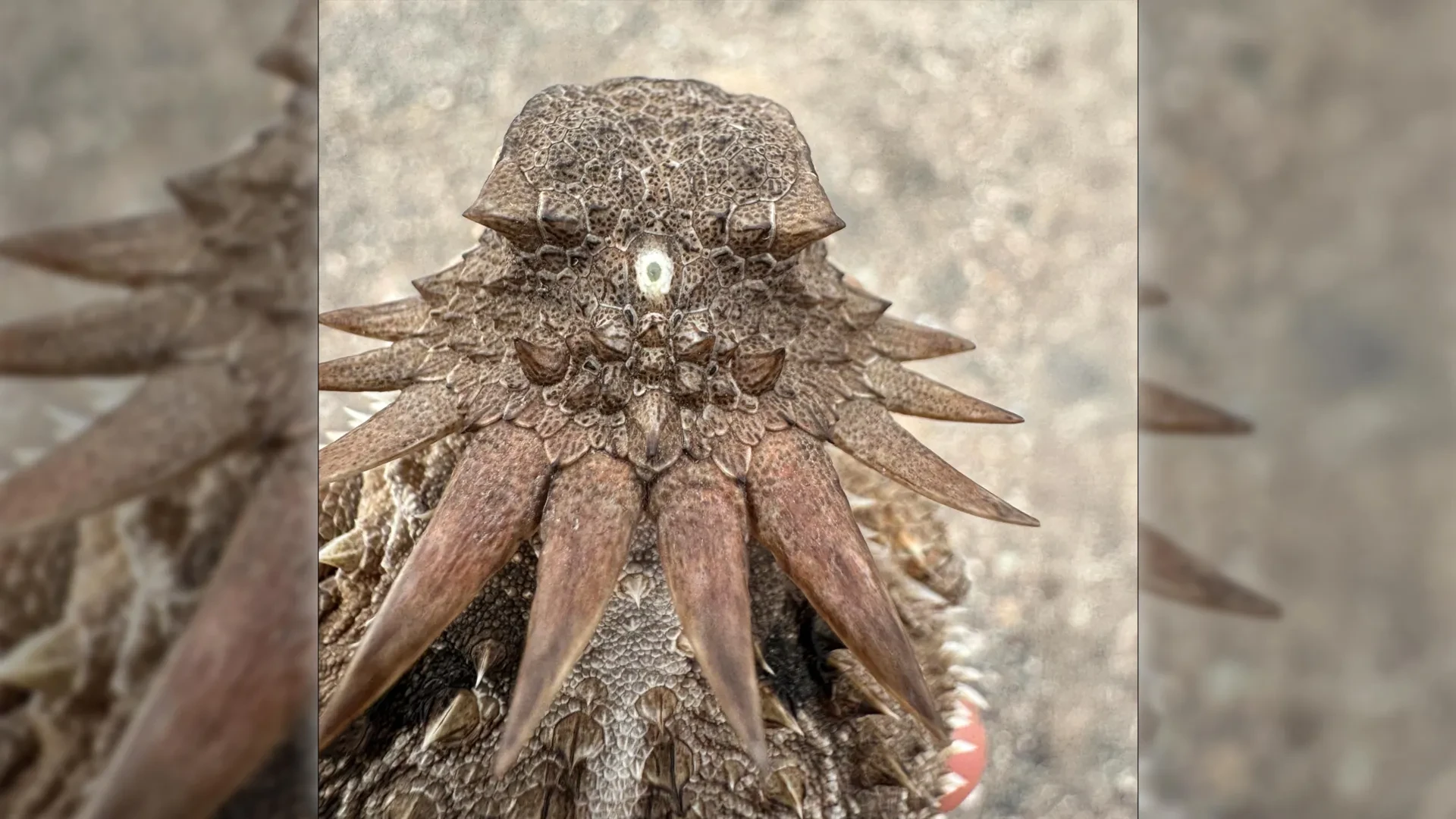
- Evolutionary Continuity: In many non-mammalian vertebrates—such as certain species of fish, amphibians, and reptiles—the pineal organ (sometimes called the parietal eye or "third eye") sits directly beneath a transparent opening in the skull, possessing a tiny lens and retina that directly register sunlight without the mediation of the primary eyes.
In mammals, the pineal gland lost its direct photoreceptive cells, but it retained its photosensitive biochemical machinery and its fundamental mandate: translating environmental light cycles into hormonal signals.
"It’s mind-boggling that our pineal gland’s ability to regulate our sleep according to light stems from the cyclopean median eye of a distant ancestor 600 million years ago," Dan-E Nilsson reflects.
4. Official Statements & Scientific Consensus
The findings, compiled through rigorous comparative genomics, neuroanatomical mapping, and evolutionary modeling by researchers at Lund University and the University of Sussex, have drawn praise and astonishment from the broader scientific community.
Dr. Aris Thorne, an evolutionary developmental biologist uninvolved in the study, remarked on the elegance of the researchers’ methodology: "For decades, the structural paradox of the vertebrate retina—being an outpocketing of the diencephalon rather than an invagination of the surface ectoderm—has been a thorn in the side of evolutionary embryologists. By demonstrating that modern vertebrate vision was effectively rebuilt from an ancient median photoreceptor after the loss of paired lateral eyes, this study provides a coherent narrative that bridges genetics, paleontology, and neuroanatomy."
The research team emphasizes that their conclusions were reached not through a single lucky fossil find, but through a meticulous synthesis of modern molecular data. By comparing the genetic toolkits that govern eye development across divergent animal phyla—including tunicates, lancelets, and jawless fish—the researchers mapped how ancient regulatory genes were co-opted, repurposed, and relocated over hundreds of millions of years.
"Our work highlights the sheer contingency and creativity of evolution," stated a co-author from the University of Sussex. "Evolution does not always move forward in a straight line. Sometimes, to build something as complex as the human visual system, nature has to take a massive detour, dismantling old systems and repurposing rudimentary organs in ways that defy our linear expectations."
5. Future Outlook: What the "Third Eye" Legacy Means for Modern Medicine and Neuroscience
As the dust settles on this paradigm-shifting discovery, neuroscientists and evolutionary biologists are already looking toward the horizon to explore the broader implications of the "cyclopean ancestor" hypothesis.
Unlocking the Secrets of Circadian Disorders
Understanding that the pineal gland is the direct evolutionary heir to an ancient median eye opens new avenues in chronobiology. By appreciating the deep ancestral roots of melatonin regulation, researchers studying circadian rhythm disorders, seasonal affective disorder (SAD), and sleep-wake cycle disruptions gain a profound appreciation for the physiological machinery governing human rest. Clinical researchers are investigating whether therapies targeting pineal function can be optimized by recognizing how deeply hardwired our light-response systems are within our evolutionary history.
Regenerative Medicine and Retinal Repair
The revelation that the vertebrate retina evolved from the brain rather than the skin offers fresh perspectives for regenerative medicine. Because retinal cells share a common neuroectodermal lineage with the central nervous system, understanding the ancient genetic switches that drove this unique developmental pathway could aid stem cell biologists. If scientists can better mimic the evolutionary and embryological cues that direct brain tissue to differentiate into complex retinal circuits, the prospect of repairing degenerative retinal diseases—such as retinitis pigmentosa and age-related macular degeneration—moves closer to reality.
A New Chapter in Evolutionary Anthropology
Ultimately, the story of our cyclopean ancestor serves as a humbling reminder of our deep biological connections to the primordial oceans. Every time we drift off to sleep under the influence of melatonin, or gaze out at the world through eyes wired directly into our brains, we are utilizing biological architecture forged 600 million years ago by a tiny, worm-like creature navigating the ancient seas with a single, glowing eye upon its head.