
Executive Overview
For generations, textbook biology taught a singular, foundational narrative regarding the dawn of multicellular life: the DNA within a newly fertilized egg was a structural "blank slate." According to this long-held dogma, the genetic material existed as a loose, chaotic, and largely unorganized bundle of chromatin. Scientists believed that this genomic soup remained unstructured and idle until the embryo reached a critical developmental milestone, woke up, and initiated its own genetic program—a process known as Zygotic Genome Activation (ZGA). Only then, textbooks suggested, did order emerge from the chaos.
That foundational narrative has now been thoroughly upended.
In a pair of landmark studies published in Nature Genetics and Nature Cell Biology, an international consortium of researchers has revealed that the genome is never a blank slate. Instead, it possesses an astonishing, highly sophisticated level of three-dimensional organization from the very earliest moments of life. Utilizing an ingenious, ultra-sensitive new mapping technology dubbed Pico-C, scientists have discovered that an elaborate 3D molecular scaffold is meticulously erected long before the genome’s master switches are flipped.
This newly discovered architecture is far from a mere structural curiosity. The spatial arrangement of DNA dictates which genes can be accessed, read, and switched on during embryogenesis—a precise choreography essential for normal cell differentiation, proper tissue development, and the prevention of congenital abnormalities and disease.
To achieve this breakthrough, the research team—led by Professor Juanma Vaquerizas and lead author Noura Maziak—first investigated rapid embryonic development in the model organism Drosophila melanogaster (the fruit fly). Their findings were staggering: rather than a period of molecular lawlessness, the hours following fertilization resemble a disciplined, highly modular construction site where DNA loops and folds with exacting precision.
Furthermore, the implications of this architecture extend far beyond developmental biology. In a companion human cell study conducted in collaboration with Professor Ulrike Kutay and her team at ETH Zürich in Switzerland, researchers demonstrated the catastrophic health consequences that occur when this architectural framework collapses. When the molecular anchors stabilizing human genome architecture are removed, cells misinterpret the structural breakdown as a viral invasion. This catastrophic false alarm triggers the innate immune system, leading to runaway inflammation and cellular pathology.
Together, these studies bridge a critical gap in our understanding of life and disease. By illuminating how the genome’s three-dimensional geometry is forged at the start of existence—and detailing the disastrous physiological fallout when that architecture fails—this research opens an entirely new frontier in molecular medicine, offering fresh avenues for diagnosing developmental disorders, targeting inflammatory conditions, and understanding the foundational mechanics of life itself.
Detailed Chronology: From Biological Dogma to the Pico-C Breakthrough
To appreciate the magnitude of the recent discoveries made by Professor Vaquerizas and his colleagues, one must trace the historical trajectory of molecular biology and the persistent technical limitations that kept the earliest moments of embryonic development shrouded in mystery.
The Era of the "Blank Slate" Dogma
For decades, cellular biologists operated under constraints dictated by available technology. When studying mammalian or invertebrate zygotes, researchers observed that transcription—the process by which RNA is synthesized from DNA templates—is largely quiescent immediately following fertilization. The maternal supplies deposited in the egg drive the earliest cell divisions.
Because measurable gene expression from the zygote’s own newly combined genome does not occur until hours (or days, depending on the species) post-fertilization, the prevailing assumption was that the physical DNA molecule must also be dormant, disorganized, and structurally unformed. Chromatin conformation capture techniques—such as Hi-C, which map how chromatin interacts across 3D space—required millions of cells to yield reliable data. Because early embryos are scarce and precious, obtaining enough material to map genomic architecture during these initial cleavage stages was practically impossible. Consequently, the prevailing "blank slate" model hardened into biological dogma.
The Innovation of Pico-C Technology
The paradigm shift began with the invention of Pico-C, a revolutionary methodology engineered to examine the three-dimensional structure of genomes with unprecedented sensitivity. Traditional chromatin conformation capture methods demand massive quantities of biological input material, making them blind to rare cell populations or the earliest, low-cell-count stages of embryogenesis.
Pico-C overcomes this bottleneck by drastically optimizing sample processing efficiency, requiring roughly ten times less material than standard Hi-C techniques. This technical leap gave scientists the high-resolution lens they needed to zoom into the nuclei of embryos during the very first hours of life, stripping away the fog of technical limitations that had obscured the pre-activation genome for decades.
Mapping the Fruit Fly Embryo
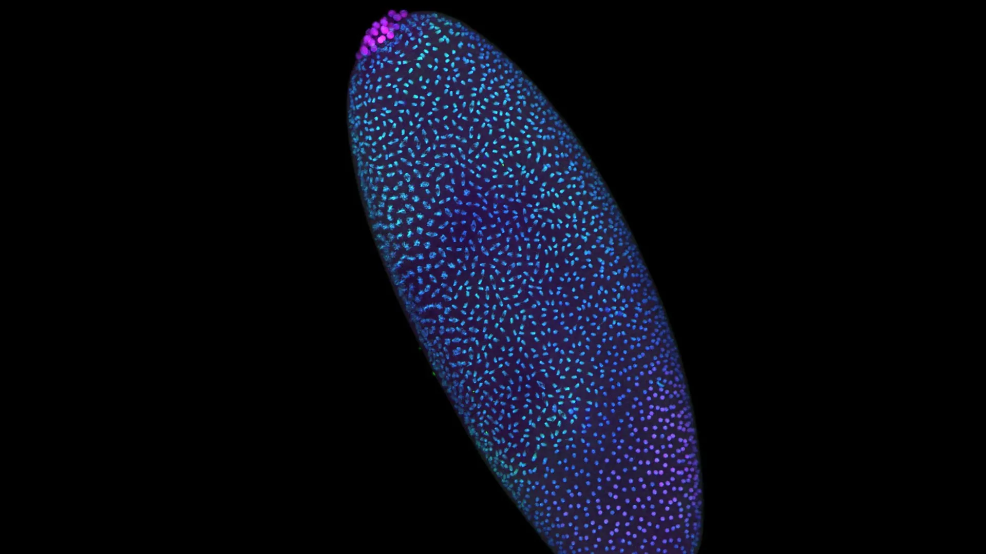
Armed with Pico-C, the research team turned to the fruit fly (Drosophila), a gold-standard model organism in genetics research. Fruit fly embryos offer a unique experimental advantage: during the first few hours following fertilization, their nuclei divide with blistering speed, producing thousands of cells in a remarkably short temporal window. This accelerated developmental pace provides an ideal laboratory ecosystem for observing how genomes assemble, organize, and regulate themselves in real-time.
Mapping the 3D chromosomal architecture of these rapidly dividing cells yielded results that stunned the research team. Long before Zygotic Genome Activation (ZGA)—the moment the embryo takes over its own genetic transcription—the DNA was not floating in a disorganized soup. Instead, it was actively folding into intricate modular loops. These spatial conformations positioned regulatory enhancers and promoters in close physical proximity, preparing the genetic machinery for activation long before the actual "on" switch was flipped.
Expanding the Lens: Human Cells and Structural Collapse
Having cracked the code of early developmental folding in fruit flies, the researchers sought to determine whether these principles applied to human biology. Collaborating with Professor Ulrike Kutay and her research group at ETH Zürich in Switzerland, the team applied high-resolution mapping strategies to human cell lines to observe what happens when genome architecture is intentionally disrupted.
Specifically, the researchers targeted the molecular "anchors"—the specialized proteins and nuclear envelope components responsible for stabilizing the genome’s intricate 3D folding patterns. When these structural anchors were experimentally degraded, the results were instantaneous and dramatic.
Deprived of its spatial scaffolding, the human genome unraveled. More alarmingly, the cell misinterpreted this structural collapse as a viral assault. The sudden exposure of naked, disorganized DNA fragments triggered the cell’s innate immune surveillance pathways, mounting an aggressive defense response against the cell’s own genetic material. This false alarm resulted in pathological inflammation, cell cycle arrest, and eventual cellular degeneration.
By connecting the precise construction of genomic architecture at life’s origin to the catastrophic consequences of its structural failure, the combined findings completed a monumental narrative arc in modern molecular biology.
Supporting Context, Metrics, and Technical Insights
Understanding the profound implications of these discoveries requires a closer look at the quantitative metrics, cellular mechanics, and technological parameters that define this research.
Quantitative Breakdown of the Research
- Sample Efficiency: Pico-C requires approximately 10 times less input material than traditional Hi-C assays, democratizing high-resolution 3D genomics for rare cell types, small tissue biopsies, and early embryos.
- Model Organism: Drosophila melanogaster yielded thousands of synchronously dividing nuclei within the first 2 to 4 hours post-fertilization, providing high temporal resolution of early genomic folding dynamics.
- Dual-Platform Validation: Findings were cross-verified across two distinct biological systems—invertebrate embryogenesis (Nature Genetics) and human cellular structural biology (Nature Cell Biology)—proving the universal evolutionary conservation of 3D genomic scaffolding.
- Funding and Institutional Support: The research was principally funded by the Medical Research Council (MRC) and the Academy of Medical Sciences (AMS), facilitated through a prestigious AMS Professorship award.
The Mechanics of 3D Genome Organization
To understand why the discovery of early genomic scaffolding is so disruptive to traditional biology, one must consider how DNA is packed inside a microscopic cellular nucleus. If stretched out straight, the DNA from a single human cell would measure roughly two meters in length. Yet, it must fit inside a nucleus just six micrometers wide—the biological equivalent of packing 40 kilometers of extremely fine thread into a tennis ball without tangling it.
Nature achieves this miracle through hierarchical folding:
- Nucleosomes: DNA is first wrapped around histone protein spools to form chromatin fibers.
- Loops and Domains: Chromatin is further folded into topologically associating domains (TADs) and chromatin loops.
- Compartmentalization: Active (euchromatin) and inactive (heterochromatin) regions segregate into distinct spatial compartments within the nuclear volume.
Before Pico-C, scientists assumed steps two and three occurred after transcription began. The new data demonstrates that the nuclear architecture is pre-engineered. The scaffolding is assembled in a modular fashion, creating a spatial map that dictates future gene expression. If a gene is required at hour four of development, its physical neighborhood is pre-configured during hour one, ensuring that transcriptional machinery can access it instantly when the developmental clock strikes the correct hour.
The Immune System’s Cryptic Warning System
The companion study from ETH Zürich provides vital context regarding the evolutionary purpose of genome architecture. The cell’s innate immune system is ancient, highly conserved, and hyper-vigilant. Its primary job is to detect foreign pathogens—specifically viral DNA and RNA—that invade the cellular cytoplasm or nucleus.
Normally, the host’s own genomic DNA is safely sequestered, neatly folded, and anchored away from cytosolic immune sensors (such as cGAS-STING pathways). However, when the 3D structural anchors fail, chromatin de-condenses, loops unravel, and chromosomal fragments leak or become abnormally exposed. The cell’s surveillance machinery mistakes these structural aberrations for viral dsDNA accumulation.
This triggers an emergency interferon-mediated immune response. In a living organism, chronic activation of this pathway by structurally compromised cells leads to sterile inflammation, autoimmune pathologies, and accelerated cellular senescence. Thus, maintaining 3D genome architecture is not merely a matter of neat housekeeping; it is a vital checkpoint distinguishing self from non-self, health from disease.
Official Statements and Expert Perspectives
The breakthrough findings have drawn widespread acclaim from the international scientific community, highlighting both the elegance of the technical achievement and the gravity of its biomedical implications.
Reflecting on the shift in perspective regarding early embryonic life, Professor Juanma Vaquerizas, principal investigator of the Nature Genetics study, noted:
"We used to look at the developmental window before the genome awakens and see a biological black box—an assumed period of chaos where structure had yet to emerge. By deploying Pico-C, we have pulled back the curtain to reveal that life begins with exquisite architectural precision. There is no chaos here; there is only masterful engineering."
Highlighting the emotional and intellectual thrill of observing these phenomena for the first time, lead author Noura Maziak described the microscopic reality of the early embryo:
"When you zoom in closer than ever before, you realize that the nucleus of a newly fertilized egg is a highly disciplined construction site. The scaffolding of the genome is being erected in a precise, modular way, long before the ‘on’ switch is fully flipped. It changes how we think about cellular memory and the transmission of developmental instructions."
Connecting the two halves of the research—the assembly of structure and the disaster of its collapse—Professor Vaquerizas synthesized the overarching thesis of the dual publications:
"These two studies tell a complete, unbroken story. The first study shows us how the genome’s 3D structure is carefully and deliberately built at the very start of life. The second study, led by our colleagues in Zurich, shows us the disastrous consequences for human health if that architectural structure is allowed to collapse."
Professor Ulrike Kutay of ETH Zürich, who spearheaded the human cell companion study published in Nature Cell Biology, emphasized the translational urgency of understanding structural anchors:
"Our work demonstrates that the physical architecture of the genome serves as a critical protective barrier. When the molecular anchors fail, the cell does not merely experience disorganization—it experiences an existential crisis, turning its own immune defenses against itself. Understanding these mechanics provides a completely new lens through which to investigate inflammatory disorders, premature aging, and degenerative human diseases."
Future Outlook: The Next Frontier in 3D Genomics
As the scientific community digests the revelations of the Pico-C methodology and its associated discoveries, the horizon of genetics research is expanding rapidly. The realization that genomic architecture precedes gene activation opens up transformative avenues of investigation across multiple disciplines.
1. Re-Evaluating Developmental Disorders and Congenital Syndromes
Many developmental abnormalities and congenital birth defects arise during the earliest hours of embryogenesis—a window that was previously difficult to interrogate due to technical constraints. With Pico-C enabling high-resolution mapping from minuscule sample sizes, researchers can now investigate whether human developmental disorders are driven not by mutated DNA sequences, but by structural misfoldings during early embryogenesis. If an embryo builds its initial 3D genomic scaffolding incorrectly, downstream gene activation will misfire, resulting in developmental failure even if the underlying genetic code is free of sequence mutations.
2. Novel Therapeutics for Inflammatory and Autoimmune Diseases
The revelation that structural collapse of the genome triggers innate immune responses via false-alarm viral sensing offers a fresh pharmacological target. By identifying the specific molecular anchors that stabilize human chromatin architecture, drug developers can design targeted therapeutics to reinforce these anchors in aging or diseased cells. Preventing structural decay could dampen pathological inflammation, mitigate autoimmune reactions, and potentially slow cellular senescence in age-related degenerative conditions.
3. Advancements in Reproductive Medicine and IVF
In assisted reproductive technologies, such as in vitro fertilization (IVF), embryologists frequently face the challenge of selecting the healthiest embryos for implantation. Current selection criteria rely heavily on morphological observations and pre-implantation genetic screening (PGS) for chromosomal number abnormalities (aneuploidy). However, many morphologically normal embryos fail to implant or develop successfully. Incorporating ultra-sensitive, low-input 3D structural profiling could eventually provide clinicians with a revolutionary biomarker for embryonic viability—assessing whether the 3D genomic construction site was set up correctly.
4. Technological Refinement and Single-Cell Resolution
As the Pico-C technology continues to evolve, researchers are pushing toward true single-cell 3D genomics during early development. While the current study maps populations of cells with unprecedented efficiency, observing cell-to-cell variability in real-time architectural folding will provide even deeper resolution into how epigenetic fate decisions are made.
Conclusion
The dogma of the genomic "blank slate" has been officially relegated to the history books. Life does not begin in structural chaos; it begins with precise, calculated architectural engineering. By pioneering the tools to visualize this hidden microscopic scaffolding—and exposing the harrowing immune consequences when that scaffolding shatters—modern researchers have charted a new map of human biology, one where space, structure, and sequence are inextricably bound in the service of life.