
Executive Overview
Deep within the evolutionary history of vertebrates—a vast lineage encompassing humans, birds, reptiles, amphibians, and fish—lies a bizarre and previously unmasked chapter. Recent groundbreaking research spearheaded by evolutionary biologists at Lund University and the University of Sussex reveals that our most distant ancestors passed through a distinctly "cyclopean" stage nearly 600 million years ago. During this archaic epoch, the precursors to modern backboned animals navigated their primordial marine environments using a single, centrally located median eye perched atop their heads.
This startling revelation upends decades of orthodox thinking regarding the evolution of sensory organs. According to the study, this ancient median eye did not merely fade into evolutionary obscurity; rather, it served as the critical biochemical and structural foundation from which our modern, paired eyes ultimately emerged. Even more astonishingly, remnants of this ancient visual apparatus persist within the human skull today: the pineal gland, a tiny endocrine organ nestled deep within the brain that regulates our circadian rhythms and sleep-wake cycles, is now understood to be the evolutionary descendant of our ancestors’ third eye.
By conducting a comprehensive, cross-species comparison of light-detecting cells, their genetic pathways, and their neural connections, the research team has solved a longstanding evolutionary puzzle. They have uncovered why vertebrate eyes are fundamentally distinct from those of invertebrates like insects and cephalopods. While a squid or a fly builds its eyes from outward-folding skin tissue, the vertebrate retina develops as an direct extension of the brain. This structural anomaly is the legacy of a strange evolutionary detour—a time when our sessile, worm-like forebears abandoned active movement, discarded their paired eyes, relied entirely on a single central eye, and later reinvented advanced vision from those very same centralized neural circuits.
Detailed Chronology: The Evolutionary Trajectory of Vertebrate Vision
To understand how humans inherited a visual system rooted in a single-eyed prehistoric organism, we must trace a timeline stretching back across six hundred million years of environmental pressures, anatomical adaptations, and genetic rewiring.
Phase I: The Primal Marine World and the Loss of Paired Eyes (Approx. 600 Million Years Ago)
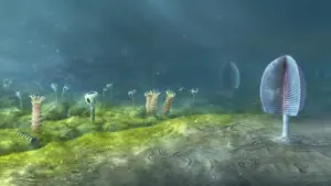
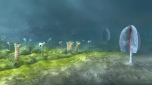
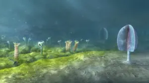
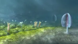
Before the Cambrian explosion truly diversified the planet’s fauna, the seas were populated by soft-bodied, microscopic pioneers. Among them was a tiny, worm-like organism that spent the majority of its existence anchored to the ocean floor. Operating as a passive filter feeder, this creature survived by sifting plankton and organic particulate matter from passing ocean currents.
In its deeper evolutionary past, this organism—or its immediate predecessors—likely possessed rudimentary paired light-sensitive cells. In the animal kingdom, paired eyes are the gold standard for mobile predators and prey alike; they allow an organism to calculate depth, gauge distances, judge trajectories, and locate external threats in a multi-dimensional space. However, for a stationary creature that rarely shifted its position, the energetic and metabolic costs of maintaining complex paired visual systems outweighed their utility.
Over countless generations, as the organism leaned further into a sedentary lifestyle, natural selection ceased to favor the maintenance of paired visual structures. Gradually, these lateral organs atrophied and disappeared altogether.
Phase II: The Rise of the Median "Cyclops" Eye
While the lateral visual fields vanished, the organism could not afford to be entirely blind to its environment. Survival in the open ocean still required a fundamental awareness of day-night cycles and spatial orientation—specifically, the ability to distinguish up from down relative to the sunlit surface.
To meet this baseline survival need, the creature retained and refined a cluster of light-sensitive cells situated precisely in the middle of its head. This yielded a primitive median eye, or "cyclopean" central organ. While incapable of resolving high-definition shapes or tracking fast-moving predators, this single central eye excelled at detecting photic gradients. It informed the organism of ambient light intensity, helping it time feeding behaviors and maintain a rudimentary vertical orientation in the water column.
Crucially, this median eye was structurally intimate with the developing neural architecture of the animal’s central nervous system. Unlike peripheral skin-derived light sensors, these cells were hardwired directly into the primordial brain tissue.
Phase III: The Great Re-Invention and the Birth of Paired Vision
As epochs shifted and ecological pressures evolved, the descendants of this sedentary worm-like creature abandoned their sedentary habits. Driven by competition for resources and the emergence of new ecological niches, these organisms returned to an active, swimming lifestyle.
A mobile existence immediately resurrected the evolutionary demand for sophisticated vision. An animal navigating a dynamic marine environment needed to detect obstacles, hunt for evasive food sources, evade predatory threats, and map its directional trajectory with precision. Yet, because their ancestors had completely lost their original lateral eyes, evolution could not simply press "rewind."
Instead, nature engineered an ingenious workaround. The organism co-opted and repurposed portions of its existing median visual structure. Through complex genetic rearrangements and developmental shifts, the central neural circuits of the median eye expanded outward, giving rise to a completely new pair of image-forming eyes.
This unusual recycling project explains a foundational quirk of vertebrate biology: why our retinas are essentially pieces of the brain pushed outward during embryonic development. When light hits our retinas today, those signals are processed by neural circuits whose ancient blueprints were originally drafted to manage input from a single, centralized median eye.
Supporting Context & Metrics: Comparative Anatomy and the Pineal Connection
To validate this evolutionary narrative, researchers had to look past superficial resemblances and examine the cellular, genetic, and neural architecture of modern species.
Vertebrates vs. Invertebrates: Two Paths to Sight
The divergence between vertebrate eyes and invertebrate eyes is one of the most striking examples of divergent evolution in biology.
- The Invertebrate Blueprint (Insects and Cephalopods): Organisms like fruit flies, bees, and squids develop their eyes from the ectoderm—the outer layer of embryonic skin tissue on the sides of the head. Their light-detecting cells (photoreceptors) utilize specific chemical cascades and signaling pathways (often rhabdomeric photoreceptors) that fold inward from the surface.
- The Vertebrate Blueprint (Humans and Mammals): In stark contrast, vertebrate eyes develop directly from the neuroectoderm—the very same embryonic tissue that forms the brain and spinal cord. Our retinal cells are ciliary photoreceptors, structurally distinct from those of insects, and are directly tethered to the central nervous system via neural pathways that process visual data before it even reaches the visual cortex.
This fundamental embryological difference has puzzled biologists for over a century. The Lund University and University of Sussex study resolves the paradox: vertebrate eyes are built this way because they were constructed from the inside out, originating from a central brain-adjacent structure rather than migrating inward from the skin.
The Pineal Gland: Living Fossil Within the Human Skull
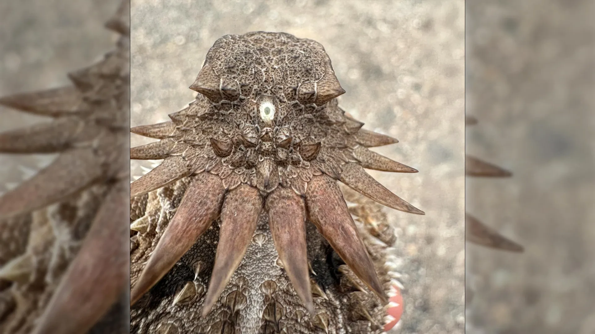
Perhaps the most poetic and tangible piece of evidence supporting this theory is the persistence of the median eye’s cellular machinery within modern vertebrates. While humans no longer possess a functional third eye on top of our skulls (though certain modern reptiles, such as the tuatara, retain a vestigial "parietal eye" complete with a lens and retina), we retain its deep interior core.
The pineal gland—a pinecone-shaped endocrine gland located in the epithalamus, tucked between the brain’s two hemispheres—is the direct evolutionary descendant of the ancient median eye.
- Melatonin Production: The pineal gland is responsible for synthesizing and secreting melatonin, a hormone that dictates the human circadian rhythm.
- Photic Regulation: In many lower vertebrates, the pineal gland remains directly photosensitive, acting essentially as a light meter. In humans, while the signal now travels indirectly via the modern paired eyes and the suprachiasmatic nucleus, the pineal gland still governs our biological clock in response to light and darkness.
This physiological reality bridges a 600-million-year evolutionary gap, linking our modern experiences of jet lag, sleep cycles, and seasonal affective responses directly to the sensory adaptations of a microscopic, single-eyed marine ancestor.
Official Statements and Expert Analysis
The implications of these findings have sent ripples through the global scientific community, prompting a re-evaluation of textbooks covering neurobiology and evolutionary developmental biology (evo-devo).
"The results are a surprise. They turn our understanding of the evolution of the eye and the brain upside down," states Dan-E Nilsson, professor emeritus in sensory biology at Lund University and a leading author of the study.
Nilsson emphasizes that mapping the lineage of light-sensitive cells required an unprecedented synthesis of comparative morphology and neurobiology. By examining where photoreceptors manifest across diverse animal phylogenies, how their cellular machinery operates, and how their nerve fibers network with neighboring tissues, the research team was able to reconstruct the sequence of events that standard fossilization processes failed to preserve. Soft-bodied organisms rarely leave behind fossil records, making this comparative cellular detective work essential.
"For the first time, we now also understand the origin of the neural circuits that analyze the image in our retina," Nilsson notes, highlighting how the internal processing units of the human eye—which handle contrast, motion detection, and brightness adjustments independently before relaying data to the brain—trace their computational lineage back to the primitive processing demands of the median eye.
Reflecting on the psychological and philosophical weight of the discovery, Nilsson adds:
"It’s mind-boggling that our pineal gland’s ability to regulate our sleep according to light stems from the cyclopean median eye of a distant ancestor 600 million years ago."
Future Outlook: Implications for Evolutionary Biology and Medicine
As the scientific community digests the conclusions put forth by the Lund and Sussex research teams, the study opens several compelling avenues for future investigation.
1. Rewriting Evolutionary Developmental Biology (Evo-Devo)
Textbooks have long categorized eye evolution through the lens of independent convergence—the idea that complex eyes evolved dozens of times across the animal kingdom from scratch. While convergence certainly plays a massive role in biology, this new research highlights the profound power of evolutionary tinkering and repurposing. Future genetic studies will likely focus on the specific transcription factors and regulatory genes (such as the Pax6 master control gene) that governed the transition from median to paired visual structures, offering deeper insights into how complex organs can be disassembled and rebuilt by natural selection over deep time.
2. Biomedical Applications in Neuro-Ophthalmology
Understanding the precise developmental link between the brain and the retina carries immense potential for regenerative medicine. Because the vertebrate retina is fundamentally an extension of the central nervous system, injuries and degenerative diseases affecting the optic nerve or retinal cells (such as retinitis pigmentosa, macular degeneration, and glaucoma) share commonalities with central nervous system disorders. By mapping the exact genetic pathways and historical lineage that forged the retina out of brain tissue, researchers gain a clearer blueprint for coaxing neural stem cells into repairing damaged ocular tissues.
3. Chronobiology and Sleep Research
On a clinical level, a deeper appreciation of the pineal gland’s ancient lineage enriches our understanding of human chronobiology. As modern society grapples with chronic sleep deprivation, blue-light exposure from digital screens, and circadian rhythm disorders, recognizing the ancient, deep-seated neurochemical pathways governing melatonin production underscores why artificial light profoundly disrupts human biology. We are attempting to trick a 600-million-year-old light-metering system designed for the open ocean with modern lighting fixtures—a mismatch that modern medicine is only beginning to fully quantify.
Ultimately, this discovery serves as a humbling reminder of our deep biological heritage. Every time a human closes their eyes to sleep, or adjusts to the morning light, they are utilizing neural architecture forged in the dim waters of the Precambrian earth—a timeless testament to the strange, brilliant, and circuitous pathways of natural evolution.