
Executive Overview
In the realm of advanced veterinary medicine, overlapping autoimmune disorders can present diagnostic puzzles that challenge even seasoned specialists. Veterinary dentistry, neurology, and internal medicine occasionally converge on a single patient whose clinical journey rewrites the textbook understanding of rare muscular pathologies. Such is the extraordinary case of Rigby, a Cavalier King Charles spaniel whose medical odyssey began at just four months of age with a severe inability to open his jaw, ultimately leading to a labyrinthine diagnostic path involving craniomandibular osteopathy (CMO), masticatory muscle myositis (MMM), and, years later, extraocular muscle myositis (EOM).
Authored from the clinical perspectives of veterinary dental and oral surgery specialist John R. Lewis, VMD, DAVDC, FF-OMFS, Rigby’s case highlights the diagnostic pitfalls inherent in pediatric canine patients presenting with trismus (restricted jaw range of motion). Initially surrendered by his original owner due to a rigid, painful jaw opening of merely 1 centimeter, Rigby was adopted by a compassionate veterinary technician who was determined to uncover the root cause of his distress.
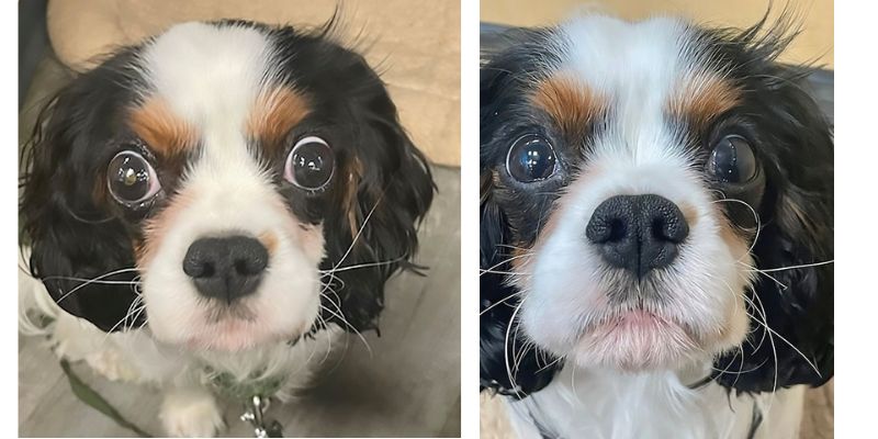
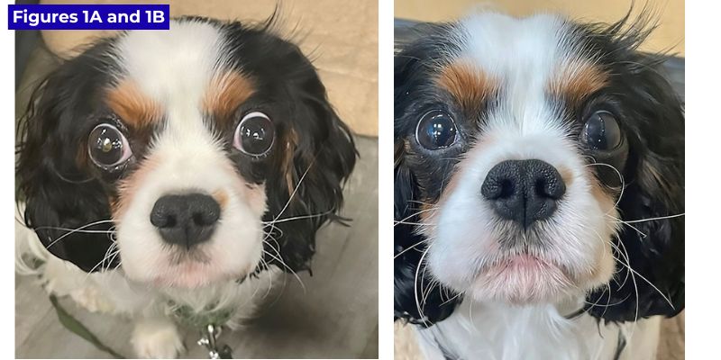
What followed was a multi-year masterclass in clinical deduction. Rigby survived canine parvovirus, lived through confounding initial computed tomography (CT) scans pointing toward early CMO, experienced a dramatic cranial structural metamorphosis, tested positive for masticatory muscle myositis, and—just when stability seemed achieved—developed acute bilateral exophthalmos (bulging eyes) driven by a completely distinct, rare autoimmune myopathy: extraocular muscle myositis.
This comprehensive report examines the chronological progression of Rigby’s case, contextualizes the physiological mechanics of type 2M muscle fibers, analyzes the diagnostic utility of advanced neuroimaging and serological assays, and explores the clinical takeaways for veterinary practitioners managing complex immune-mediated canine myopathies.
Detailed Chronology of a Medical Odyssey
The Early Presentation: Trismus and the Initial Surrender
Rigby entered the veterinary landscape at four months of age as a vulnerable puppy unable to interact normally with his environment due to a locked jaw. With an interincisal distance (the vertical gap between the upper and lower front teeth) limited to just 1 centimeter, the young Cavalier King Charles spaniel demonstrated clear signs of pain during any physical manipulation intended to increase his jaw range of motion. Recognizing the severity of the condition and the long-term commitment required for management, his original owner surrendered him. Fortunately, the puppy captured the heart of a veterinary technician who formally adopted him, providing the stable home environment and vigilant observation necessary to track his unfolding medical chapters.
[4 Months Old] ──> Severe Trismus (1cm gap) ──> Surrender & Adoption by Vet Tech
      │
      ├──> CT Scan: TMJ Dysplasia & Early CMO Suspected
      └──> Intercurrent Illness: Canine Parvovirus (Jaw opens to 1.3cm)
The Diagnostic Labyrinth: CT Scans, CMO, and Parvovirus
Shortly after adoption, Rigby underwent a comprehensive computed tomography (CT) scan evaluated by a board-certified veterinary radiologist. The imaging revealed:
- Bilateral temporomandibular joint (TMJ) dysplasia.
- Bilateral TMJ subluxation accompanied by a widening of the right temporomandibular joint space.
- Scant periosteal new bone formation along the left mandibular ramus, situated ventrally to the condyloid process.
- Bilateral thickening of the walls of the rostral tympanic bullae.
- Normal calvarium appearance, with no mineralized bony bridges connecting the TMJ structures to the tympanic bullae.
Based on these specific bone-remodeling characteristics, the radiologist interpreted the CT findings as highly suggestive of early craniomandibular osteopathy (CMO)—a proliferative bone disease classically seen in young growing terriers and large-breed dogs, though occasionally reported in spaniels.
Almost concurrently, Rigby contracted canine parvovirus. During this systemic crisis, his jaw mobility slightly expanded to 1.3 centimeters. Once he successfully conquered the parvovirus and recovered his strength, Rigby was referred to Dr. Lewis’s specialty service for definitive management of his suspected CMO and TMJ limitations.
A Surprising Metamorphosis and the Pivotal Shift in Diagnosis
Upon presentation to the dental and oral surgery service, Rigby was remarkably thriving at home. He ingested both wet and dry commercial dog food with enthusiasm, displaying no systemic malaise. However, his pet parent noted a striking physical transformation: the puppy’s head, which previously appeared abnormally broad, flat, and swollen on top, had gradually evolved into a more normal, dome-shaped Cavalier conformation.
This clinical observation acted as the critical diagnostic catalyst for Dr. Lewis. While transient head flattening and subsequent reshaping could theoretically be attributed to disuse muscle atrophy during his prolonged period of restricted jaw movement, another distinct disease process immediately came to mind: masticatory muscle myositis (MMM).
[Recovery Phase] ──> Head Conformation Change (Swollen to Domed) ──> Suspicion of MMM
      │
      ├──> Physical Exam: 3.8cm Jaw ROM, Temporal/Masseter Atrophy
      └──> 2M Antibody Test: Positive (1:1000 Titer) ──> Diagnosed with MMM
Physical examination revealed:
- Normal bilateral retropulsion of the eyes.
- Moderate, symmetrical atrophy of the temporal and masseter muscles.
- A dramatic improvement in jaw range of motion, measuring 3.8 centimeters interincisal distance.
- Complete absence of pain upon repeated opening and closing of the jaws.
- Minimal side-to-side jaw laxity, rendering congenital TMJ dysplasia far less likely as the primary driver.
Confirming Masticatory Muscle Myositis (MMM)
To investigate the MMM hypothesis, blood samples were dispatched to the Comparative Neuromuscular Laboratory at the University of California, San Diego, for the specialized 2M antibody assay.
While classic MMM presentations routinely manifest with severe acute pain when owners or clinicians attempt to force the mouth open, chronic phases or waxing-waning clinical courses can mask this acute distress. Rigby’s serology returned a stellar positive titer of 1:1000, unequivocally confirming masticatory muscle myositis.
The initial treatment protocol prescribed an immune-modulating regimen of prednisolone at 0.55 mg/kg every 12 hours for seven days, followed by 0.55 mg/kg once daily for one month, with a subsequent slow taper. However, because Rigby’s internal medicine specialist and owner noted that he was clinically vibrant and asymptomatic at home, systemic corticosteroids were ultimately withheld. In a testament to the unpredictable nature of immune-mediated diseases, a repeat 2M antibody titer performed three months later plummeted to less than 1:100, and Rigby maintained a completely normal, pain-free range of jaw motion.
The Second Act: The Emergence of Extraocular Myositis
Peace reigned for two years. Rigby matured into an affectionate, healthy two-year-old spaniel with full functional mobility. However, a brand-new medical mystery emerged. Without warning, Rigby developed bilateral exophthalmos—a noticeable, symmetrical forward displacement of the globes (bulging eyes).
[2 Years Later] ──> Acute Bilateral Exophthalmos ──> Differential Diagnoses Formulated
      │
      ├──> MRI Evaluation: Atrophied Masticatory Muscles, Normal Salivary Glands
      ├──> Clinical Deduction: Extraocular Muscle Myositis
      └──> Treatment: Prednisolone (0.8 mg/kg BID) ──> Rapid, Dramatic Resolution
While acute inflammation tied to recurring or smoldering MMM can occasionally trigger bilateral exophthalmos, differential diagnoses for sudden-onset globe protrusion in young dogs also include:
- Sialadenitis of the zygomatic salivary glands.
- Primary extraocular muscle myositis (EOM).
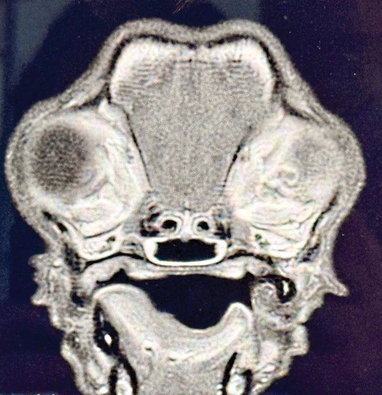
An advanced Magnetic Resonance Imaging (MRI) scan was commissioned to map the soft tissue architecture of the retrobulbar and cranial spaces in high resolution. The evaluating radiologist noted an absence of inflammation in the masticatory muscles—confirming persistent, quiescent atrophy from his prior MMM episode. Furthermore, the zygomatic salivary glands beneath the eyes appeared completely normal in size and symmetry, and the retrobulbar space showed no gross structural asymmetry.
Upon re-evaluating the MRI alongside the radiologist’s notes, Dr. Lewis deduced that the extraocular muscles were the most probable anatomical source of the exophthalmos. By process of elimination—ruling out active masticatory inflammation and salivary gland pathology—extraocular muscle myositis stood as the definitive culprit.
Rigby was promptly initiated on a therapeutic trial of prednisolone at 0.8 mg/kg twice daily. The response was nothing short of cinematic: rapid, dramatic regression of the globe protrusion and restoration of normal ocular alignment. He was subsequently transitioned to a long, ultra-slow corticosteroid taper and currently maintains complete clinical remission on a low maintenance dose of 0.2 mg/kg of prednisolone once daily.
Supporting Context & Pathophysiological Metrics
To fully appreciate the complexity of Rigby’s medical journey, one must examine the distinct cellular biology governing canine cranial musculature.
The Unique Biology of Type 2M Muscle Fibers
Masticatory muscle myositis is an idiopathic autoimmune inflammatory myopathy whose clinical manifestations are strictly anatomically restricted. The primary muscles responsible for jaw closure—specifically the temporal, masseter, and medial and lateral pterygoid muscles—are embryologically and biochemically distinct from skeletal muscles located throughout the rest of the canine body.
These masticatory muscles are exclusively composed of type 2M muscle fibers, which contain a unique, specialized isoform of myosin heavy chains found nowhere else in the somatic musculaturer. Conversely, the digastricus muscle (responsible for opening the jaw) is composed of standard type 2A fibers and remains entirely unaffected by the disease process.
+---------------------------+-----------------------------------+-----------------------------+
| Muscle Group              | Fiber Type                        | Susceptibility to MMM       |
+---------------------------+-----------------------------------+-----------------------------+
| Temporal                  | Type 2M                           | Highly Susceptible          |
| Masseter                  | Type 2M                           | Highly Susceptible          |
| Medial/Lateral Pterygoid  | Type 2M                           | Highly Susceptible          |
| Digastricus               | Type 2A                           | Immune / Unaffected         |
| Extraocular Muscles       | Specialized Myosin Isoforms       | Susceptible to EOM Variant  |
+---------------------------+-----------------------------------+-----------------------------+
When a dog develops MMM, circulating autoantibodies specifically target these type 2M fibers. This targeted humoral and cell-mediated immune response prompts a massive infiltration of inflammatory cells (lymphocytes, plasma cells, and macrophages) into the muscle bellies. The resulting immune complex formation drives localized tissue necrosis, active phagocytosis, and eventual fibrous connective tissue replacement (fibrosis), which clinically manifests as either acute pain and trismus or chronic muscle wasting and end-stage contracture.
Diagnostic Precision: Serology, Imaging, and Histology
The commercially available serum 2M antibody test represents a monumental diagnostic breakthrough in veterinary neurology. It boasts an exceptional sensitivity rating of 85% to 90% and a specificity approaching 100% for classical masticatory muscle myositis.
However, clinicians must exercise extreme caution regarding timing: administering immunosuppressive corticosteroid therapy within 10 days prior to blood collection can dramatically blunt antibody production, yielding a false-negative titer.
When serology is ambiguous or prior steroid administration muddies the waters, advanced imaging and histopathology provide definitive answers:
- Computed Tomography (CT): While valuable for ruling out osseous changes (such as CMO or TMJ ankylosis), conventional CT scans in MMM cases often reveal patchy, heterogeneous contrast uptake patterns reflecting regional variations in muscular inflammation.
- Magnetic Resonance Imaging (MRI): Unrivaled for soft-tissue resolution, MRI allows clinicians to differentiate between atrophied masticatory bellies, normal or enlarged salivary structures, and swollen retrobulbar or extraocular tissues.
- Muscle Biopsy: Surgical sampling of the temporal muscle followed by immunohistochemical staining remains the gold standard. It reveals classic immune complex deposition surrounded by intense inflammatory cuffs, confirming the diagnosis even in cases where serum antibody titers are temporarily suppressed.
Comparative Literature and Breed Predispositions
Rigby’s case sits at the intersection of two exceedingly rare veterinary conditions: atypical juvenile masticatory muscle myositis and extraocular muscle myositis.
Juvenile Atypical MMM in Cavalier King Charles Spaniels
Classical MMM typically affects young adult to middle-aged large-breed dogs (such as German Shepherds, Doberman Pinschers, and Golden Retrievers). However, pediatric presentations in toy breeds are documented in veterinary literature. A landmark case series detailed an atypical presentation of juvenile MMM occurring within a single litter of Cavalier King Charles spaniels. In that report, three out of four 12-week-old littermates presented with severe trismus and difficulty opening the mouth. Diagnosis was successfully established via 2M immunohistochemistry and characteristic temporal muscle histopathology, and all affected puppies achieved complete clinical resolution following prompt implementation of immune-modifying corticosteroid therapy. Rigby’s early-life trismus mirrors this rare familial predisposition.
Extraocular Muscle Myositis (EOM)
Extraocular muscle myositis is an exceptionally uncommon entity in peer-reviewed veterinary literature. A foundational case series reviewing 10 young dogs documented presentations that were predominantly bilateral or unilateral across varied canine breeds. Clinically, abnormalities were strictly restricted to the extraocular muscles, leaving the masticatory and appendicular skeletal muscles entirely unaffected. Histological examination of EOM cases characteristically reveals variable lymphocytic-plasmacytic mononuclear cell infiltration alongside interstitial fibrosis.
Furthermore, rare case reports have documented variants of extraocular myositis driven explicitly by eosinophilic inflammation. This carries profound immunological significance for Rigby’s specific breed: Cavalier King Charles spaniels are already well-documented in veterinary medicine to possess a genetic predisposition toward broader eosinophilic disease manifestations, including eosinophilic palatitis (eosinophilic stomatitis of the palate). Although Rigby’s exact cellular subtype in his extraocular flare remains clinically managed via empirical steroid responsiveness, the breed propensity for immune-mediated eosinophilic and muscular reactivity underscores the interconnected nature of his systemic health history.
Future Outlook & Clinical Takeaways
Rigby’s triumphant management—navigating neonatal trismus, parvoviral infection, presumptive CMO, confirmed masticatory muscle myositis, and secondary extraocular myositis—serves as an inspiring testament to modern veterinary diagnostics and the unwavering dedication of a veterinary technician pet parent.
As Rigby continues to thrive under his long, slow maintenance protocol of low-dose prednisolone (currently stable at 0.2 mg/kg once daily), his case leaves several enduring lessons for veterinary practitioners:
- Broaden the Differential for Puppy Trismus: When a juvenile dog presents with restricted jaw mobility, clinicians must look beyond congenital joint dysplasias and developmental bone disorders (such as CMO) to consider autoimmune myopathies, including atypical juvenile MMM.
- Recognize Conformation Shifts: Sudden alterations in cranial conformation—such as the transition from a swollen, flat head to a normal dome shape—can serve as vital clinical clues pointing toward subacute masticatory muscle atrophy.
- Anticipate Multi-System Autoimmunity: Dogs diagnosed with one organ-specific or muscle-specific immune-mediated myopathy may harbor predispositions to related immunological flare-ups later in life, necessitating vigilant long-term monitoring of ocular, orbital, and masticatory structures.
- Leverage Advanced Imaging and Interdisciplinary Collaboration: The synthesis of high-resolution MRI, precise serological testing (2M titers), and collaborative peer review between dental specialists, radiologists, and internal medicine clinicians remains the ultimate formula for unraveling complex canine autoimmune diseases.
Rigby stands today as a living example of how clinical vigilance, advanced imaging, and tailored immunomodulatory therapy can rescue a patient from the brink of diagnostic confusion, granting them a vibrant, comfortable, and pain-free life.
References
- Shelton, G. D. (2014). Canine Inflammatory Myopathies: Masticatory Muscle Myositis, Extraocular Muscle Myositis, and Polymyositis. Veterinary Clinics of North America: Small Animal Practice, 44(4), 779-797.
- Evans, J., et al. (2005). Atypical Masticatory Muscle Myositis in a Litter of Cavalier King Charles Spaniels. Journal of Veterinary Internal Medicine, 19(3), 395-399.
- Gilger, B. C., et al. (2002). Extraocular Muscle Myositis in Dogs: 10 Cases (1990-2000). Journal of the American Veterinary Medical Association, 220(10), 1503-1507.
- Pfeiffer, C., et al. (2011). Eosinophilic Extraocular Myositis in a Dog. Veterinary Ophthalmology, 14(2), 125-130.
- Craig, L. E., et al. (2008). Eosinophilic Stomatitis and Palatitis in Cavalier King Charles Spaniels. Veterinary Pathology, 45(3), 346-351.