
Executive Overview
Pyometra—a severe, life-threatening infection of the uterus—remains one of the most critical reproductive emergencies encountered in small animal veterinary medicine. Left unrecognized or untreated, the condition rapidly progresses from localized uterine inflammation to systemic inflammatory response syndrome (SIRS), endotoxemia, multiple organ dysfunction syndrome (MODS), and death.
This clinical case study examines the presentation, diagnostic evaluation, pathophysiological mechanisms, and risk factor profile of a nine-year-old intact female Bulldog evaluated by Kendra Freeman, DVM, MS, DACVS. The patient presented with classic, albeit non-specific, clinical signs of systemic illness: lethargy, hyporexia, dehydration, and acute abdominal pain. Laboratory diagnostics revealed marked leukocytosis characterized by a neutrophilia, concurrent azotemia indicative of renal compromise, and hyperglobulinemia reflecting chronic antigenic stimulation.
Beyond evaluating the individual patient, this report explores the broader epidemiological and endocrinological drivers of canine pyometra. By analyzing breed predilections, hormonal influences such as estrogen and progesterone exposure, and the statistical likelihood of disease development in intact female dogs, this article provides veterinary professionals and advanced students with an authoritative, comprehensive review of canine reproductive pathology.
Detailed Chronology: Patient Presentation and Clinical Findings
Initial Presentation and Anamnesis
The patient, a nine-year-old intact female Bulldog, was brought to the veterinary clinic with a multi-day history of progressive lethargy, reluctance to move, and a significant decrease in appetite. According to the owner, the dog had become increasingly apathetic, spent the majority of her time recumbent, and showed zero interest in her standard diet. No overt vaginal discharge was initially reported by the owner, a common presentation in "closed" pyometra cases where the cervix remains tightly sealed, preventing the external escape of purulent exudate.
Physical Examination Findings
Upon presentation, Dr. Kendra Freeman performed a comprehensive physical examination. The patient’s vital signs and general physical parameters revealed several critical abnormalities:
- Mucous Membranes: Tacky to dry, indicating moderate-to-severe systemic dehydration secondary to decreased fluid intake, ongoing fluid sequestration within the uterine lumen, and potential renal fluid loss.
- Abdominal Palpation: Tense and painful upon deep palpation of the caudal and mid-abdomen. The discomfort localized primarily to the ventral midline and retroperitoneal spaces, consistent with a fluid-distended, inflamed uterus exerting pressure on surrounding visceral organs.
- Cardiorespiratory System: Tachycardia commensurate with systemic inflammation, dehydration, and pain. Pulses were adequate, but capillary refill time was marginally prolonged at approximately 2.5 seconds.
Diagnostic Workup and Laboratory Findings
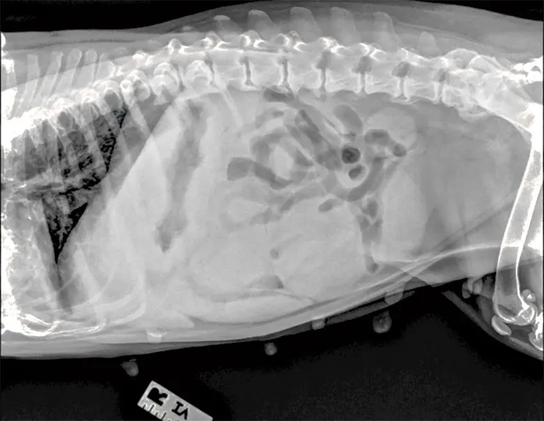
To isolate the underlying etiology of the patient’s systemic decline, a complete blood count (CBC), serum biochemistry profile, and diagnostic imaging (abdominal radiography) were immediately initiated.
-
Complete Blood Count (CBC):
- Marked leukocytosis characterized by a profound neutrophilia, frequently accompanied by a left shift (regenerative or degenerative depending on bone marrow reserve). This hematologic pattern is the hallmark of a severe, acute bacterial infection and intense systemic inflammation. Toxic changes within neutrophils (such as Döhle bodies and cytoplasmic basophilia) are frequently observed in these cases, pointing toward accelerated granulopoiesis driven by circulating inflammatory cytokines (e.g., IL-1, IL-6, TNF-alpha).
-
Serum Biochemistry Profile:
- Azotemia: Elevations in both blood urea nitrogen (BUN) and creatinine were identified. This finding typically points toward prerenal azotemia secondary to severe dehydration, though concurrent renal injury induced by circulating bacterial endotoxins (tubular nephrosis or glomerulonephritis) is a well-documented complication of canine pyometra.
- Hyperglobulinemia: Elevated serum globulin fractions reflected chronic antigenic stimulation and systemic activation of the humoral immune response. As the uterus fills with bacteria, bacterial toxins, and cellular debris, constant systemic absorption drives hepatic synthesis of acute-phase proteins and immunoglobulin production.
-
Diagnostic Imaging (Abdominal Radiography):
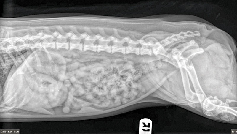
- Lateral abdominal radiographs revealed a prominent, tubular soft-tissue tubular opacity within the mid-to-caudal abdomen, displacing the intestinal loops cranially and dorsally. This radiographic appearance is pathognomonic for a fluid-filled, enlarged uterine structure in an intact adult female canine.
Supporting Context & Metrics: Epidemiology, Pathophysiology, and Risk Factors
To contextualize this case, veterinary practitioners must understand the underlying risk factors, incidence rates, and microbiological profiles associated with canine pyometra.
1) Risk Factors for the Development of Pyometra
The development of pyometra is intimately linked to the hormonal milieu of the canine estrous cycle, specifically the prolonged luteal phase characteristic of the canine species.
- Estrogen Administration and Exposure: Exogenous estrogen administration (historically used for mismatched breeding termination) sensitizes the canine endometrium by upregulating progesterone receptors. When followed by endogenous progesterone secretion during metestrus, the endometrium undergoes exaggerated proliferation and glandular secretion.
- Increased Age: The cumulative risk of pyometra increases exponentially with age. Each successive estrous cycle exposes the feline or canine endometrium to repeated bouts of progesterone-induced cystic endometrial hyperplasia (CEH). Consequently, middle-aged to older intact females (typically >6 years of age) represent the classic demographic.
- Breed Predispositions: While pyometra can affect any intact female dog, certain breeds exhibit a markedly elevated genetic predisposition. Breeds frequently cited in veterinary literature include Golden Retrievers, Miniature Schnauzers, Rottweilers, Cavalier King Charles Spaniels, and Bulldogs.
Correct Answer to Clinical Challenge Question #1: d) All of the above (Retriever breeds, estrogen administration/exposure, and increased age are all established, clinically significant risk factors for the development of canine pyometra).
2) The Epidemiological Incidence of Pyometra
Pyometra is not a rare anomaly; rather, it is one of the most prevalent reproductive disorders in intact female dogs. Epidemiological studies tracking intact female populations demonstrate that a substantial percentage of bitches will develop the condition if they remain unspayed throughout their lifespan.
Landmark veterinary epidemiological investigations have consistently shown that approximately one in four intact female dogs will develop pyometra by 10 years of age. Certain high-risk breeds see incidence rates climb even higher.
Correct Answer to Clinical Challenge Question #2: c) 25 percent (Statistical analyses indicate that approximately 25%—or one-fourth—of all intact female dogs will develop pyometra within their lifetimes).
3) Microbiological Profile of Uterine Discharge
The pathogenesis of pyometra begins with hormonal priming (CEH), which creates an immunologically suppressed, nutrient-rich environment within the uterine lumen. Under these conditions, normal ascending bacterial flora from the vagina—or hematogenous seeding—can easily colonize the tissue.
While multiple opportunistic pathogens can be isolated from infected uterine horns, one specific organism dominates the microbiological landscape of canine pyometra due to its unique virulence factors, including fimbriae that adhere specifically to canine endometrial receptors.
Correct Answer to Clinical Challenge Question #3: a) E. coli (Escherichia coli is overwhelmingly the most common bacterial isolate recovered from pyometra discharge, accounting for the vast majority of clinical cases worldwide).
Official Statements and Expert Perspectives
The management of pyometra requires a standardized, highly aggressive protocol blending emergency stabilization with definitive surgical intervention. Leading veterinary surgeons and academicians emphasize that pyometra should never be treated as an elective outpatient procedure.
Dr. Kendra Freeman, the managing clinician on this case, underscores the urgency required when dealing with geriatric intact patients presenting with vague signs of systemic malaise:
"When an intact, middle-aged or older female dog presents with lethargy, a tense abdomen, and subclinical or overt signs of dehydration, pyometra must immediately sit at the top of the differential diagnosis list—even in the absence of a visible vaginal discharge. The clinician’s window to prevent systemic inflammatory response syndrome and irreversible renal failure is remarkably narrow. Ovariohysterectomy is not merely a routine elective spay in these patients; it is a critical, life-saving source-control procedure."
Veterinary critical care specialists further elaborate on the physiological management of these compromised patients:
"Stabilization before and during surgery is just as vital as the surgical excision of the infected uterus. Aggressive intravenous fluid therapy, broad-spectrum antimicrobial administration targeting gram-negative anaerobes and aerobes, and continuous monitoring of acid-base status and renal parameters dictate whether a patient survives the immediate postoperative period."
Future Outlook: Prevention, Early Detection, and Surgical Innovations
The Imperative of Canine Ovariohysterectomy
The ultimate future outlook for eliminating pyometra as a mortality threat in companion animals lies in proactive preventive medicine. Early elective ovariohysterectomy (spaying before the first or second estrous cycle) remains the gold standard of preventative veterinary medicine. By removing the primary source of estrogen and progesterone production, the cascade of cystic endometrial hyperplasia and subsequent bacterial colonization is entirely prevented.
Emerging Medical Management Alternatives
While surgical ovariohysterectomy remains the undisputed standard of care for pets with closed or open pyometra, medical management strategies are occasionally explored under strict clinical criteria. These protocols are typically reserved for valuable breeding animals where future reproductive capability is deemed mandatory by the owner.
Medical protocols generally combine:
- Progesterone Receptor Antagonists / Prostaglandins: Agents such as aglepristone (where available) or prostaglandin F2-alpha (PGF2a) analogues are utilized to induce luteolysis, relax the cervix, and expel uterine purulent material.
- Targeted Antimicrobial Therapy: Guided strictly by uterine culture and sensitivity testing.
- Intensive Supportive Care: Fluid resuscitation, electrolyte balancing, and serial ultrasonographic monitoring of uterine involution.
However, veterinary consensus panels continually warn that medical management carries significantly higher risks of treatment failure, uterine rupture, peritonitis, and recurrence during subsequent estrous cycles. Consequently, surgical intervention remains the definitive recommendation across all global veterinary standards of care.
Diagnostic Advancements in Early Screening
Looking ahead, veterinary researchers are focusing on early biomarker identification to catch pyometra before clinical signs become overt. Advanced assays tracking acute-phase proteins (such as C-reactive protein and serum amyloid A) combined with point-of-care reproductive tract ultrasonography are expected to shorten the diagnostic timeline for primary care veterinarians. By identifying subclinical endometrial changes and mild fluid accumulation early in the diestrual phase, practitioners can intervene before patients reach the severe, life-threatening metabolic crises seen in cases like this nine-year-old Bulldog.
Conclusion
The presentation of a nine-year-old intact female Bulldog with lethargy, dehydration, abdominal pain, leukocytosis, and azotemia serves as a textbook clinical scenario of canine pyometra. Through rigorous physical examination, comprehensive hematological and biochemical profiling, and prompt surgical intervention, veterinary professionals successfully navigate these complex emergency cases. By understanding the underlying epidemiological data—including the 25% lifetime incidence rate, the predominance of E. coli infections, and the compounding risk factors of age and hormonal exposure—the veterinary community continues to refine diagnostic accuracy and advocate for proactive spay programs that safeguard canine health worldwide.