
Executive Overview
For decades, modern embryology and molecular genetics rested on a foundational assumption: the DNA inside a newly fertilized egg was a structural "blank slate." Conventional biological dogma held that the early embryonic genome existed as a loose, disorganized, and chaotic tangle of chromatin. According to this long-standing narrative, the genetic material remained largely unstructured until the embryo underwent Zygotic Genome Activation (ZGA)—the critical milestone when the embryo initiates its own genetic program and "wakes up" to direct development. Until that point, scientists believed, the genome was inert and structurally formless.
A pair of landmark studies published in Nature Genetics and Nature Cell Biology shatters this decades-old paradigm. Spearheaded by Professor Juanma Vaquerizas and his research team, alongside a companion collaboration led by Professor Ulrike Kutay at ETH Zürich in Switzerland, scientists have unveiled a breathtaking reality: the genome is not chaotic at the dawn of life. Instead, it exhibits an astonishingly complex and precise level of three-dimensional organization long before the first genes are ever transcribed.
This monumental discovery was made possible by the invention of Pico-C, a groundbreaking, ultra-sensitive chromosome conformation capture technology. Pico-C allows researchers to examine the spatial folding and 3D architecture of genetic material with unprecedented resolution, requiring a fraction of the biological material previously necessary. Using this technology in fruit fly (Drosophila) models and human cells, the research teams demonstrated that an intricate, modular DNA scaffold is meticulously erected during the earliest hours of embryonic development.
Furthermore, the companion study in human cells illuminated the catastrophic stakes of this architecture. When the molecular anchors stabilizing this 3D framework are compromised, human cells mistakenly perceive the structural collapse as a viral invasion. This misinterpretation triggers an inappropriate innate immune response, sparking chronic inflammation and pathological disease. Together, these findings bridge a critical knowledge gap in developmental biology, reframing the pre-activation embryo from a disorganized cellular soup into a highly disciplined, exquisitely coordinated construction site.
Detailed Chronology of the Scientific Breakthrough
To appreciate the magnitude of the recent discoveries by Professor Vaquerizas, Dr. Noura Maziak, Professor Kutay, and their international colleagues, it is essential to trace the historical trajectory of genomics and the technological limitations that kept the pre-activation genome shrouded in mystery.
The Dogma of the Blank Slate
Since the late 20th century, molecular biologists understood that maternal mRNAs and proteins deposited inside the egg drove the earliest divisions of a zygote. Because the paternal and maternal pronuclei fuse and undergo rapid cellular divisions without immediately transcribing new zygotic RNA, geneticists inferred that chromatin architecture must be similarly idle. Standard imaging techniques and early chromatin conformation capture methods—such as Hi-C—lacked the sensitivity to probe tiny, early-stage embryos. Consequently, textbooks taught that chromatin organization was established after genome activation, emerging as a downstream consequence of transcription rather than its prerequisite.
The Innovation of Pico-C Technology
The turning point arrived when Professor Vaquerizas’s laboratory set out to bypass the severe sample-size constraints of traditional genomic assays. Standard methods require millions of cells to map spatial DNA folding interactions reliably, making it virtually impossible to study rare, early-stage embryos or precious biopsy samples.
By refining molecular biology protocols and proximity ligation techniques, the team engineered Pico-C. This revolutionary technology scales down the required input material by a factor of ten, enabling high-resolution 3D genome mapping from microscopic biological samples. The development of Pico-C effectively opened a molecular window into the previously invisible developmental hours immediately following fertilization.
Mapping the Fruit Fly Embryo
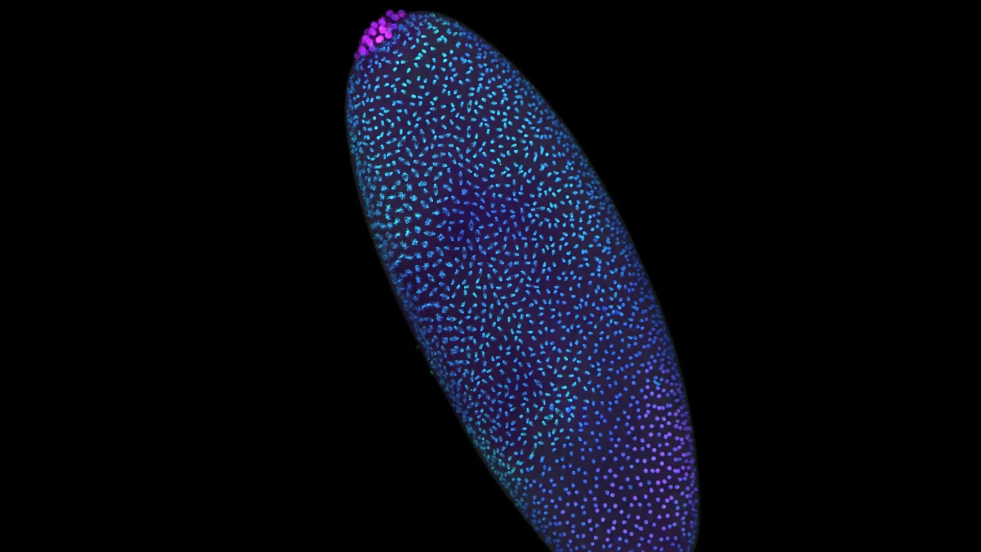
Turning to the fruit fly (Drosophila melanogaster)—a classical workhorse of genetic research renowned for its rapid embryonic development—the researchers applied Pico-C to map chromatin folding across time. In the fruit fly, the early embryo undergoes rapid nuclear divisions within a shared cytoplasm, producing thousands of nuclei in a matter of hours. This accelerated developmental timeline makes Drosophila an ideal experimental crucible.
Using Pico-C, the team tracked chromatin organization during these frantic initial hours. To their astonishment, the data revealed that DNA does not drift randomly. Instead, it folds into distinct, modular loops and spatial domains long before Zygotic Genome Activation. This physical pre-patterning ensures that regulatory elements—enhancers and promoters—are positioned in proximity to their target genes, primed and ready to fire the exact millisecond the genetic program switches on.
Unraveling Human Structural Collapse
While the Drosophila study unveiled how the 3D scaffold is constructed, a critical question remained: What happens if this architectural blueprint fails, particularly in human systems?
To answer this, Professor Vaquerizas teamed up with Professor Ulrike Kutay and her research group at ETH Zürich. In a companion study published in Nature Cell Biology, the Swiss-led team applied high-resolution mapping strategies to human cells, specifically investigating the molecular anchors responsible for maintaining nuclear architecture and chromatin folding.
When the researchers experimentally dismantled these structural anchors, the consequences were immediate and dramatic. The human genome’s spatial framework unraveled, causing DNA loops to collapse into disordered heaps. More alarmingly, the cell’s surveillance machinery misinterpreted this structural disintegration as an aggressive viral takeover. The innate immune system sounded a false alarm, triggering inflammatory pathways that mimic pathogenic infection and drive cellular pathology.
Supporting Context, Methodology, and Metrics
The implications of these findings extend far beyond basic embryology, touching upon biophysics, immunology, and the etiology of congenital disorders and degenerative diseases.
The Biomechanics of DNA Folding
DNA is an immensely long polymer. In a human cell, if stretched out, it would measure roughly two meters, yet it must pack neatly into a microscopic nucleus measuring only a few micrometers across. Achieving this compaction without tangling requires sophisticated spatial organization.
Topologically Associating Domains (TADs) and chromatin loops dictate which genetic regions interact with one another. In the traditional view of early development, these domains were thought to crystallize only after transcription factors began binding to active genes. The Pico-C data proves the inverse: the physical folding precedes and enables correct gene regulation. Space dictates function. By pre-assembling the genomic scaffold, the cell ensures that developmental master switches are thrown in the correct sequence, preventing transcriptional misfires that could lead to developmental arrest or embryonic lethality.
Pico-C: Technical Superiority and Metrics
The deployment of Pico-C represents a methodological leap forward in spatial genomics. Traditional Hi-C protocols demand substantial quantities of starting material—frequently millions of cells—to generate statistically robust contact matrices. This requirement creates a prohibitive bottleneck for:
- Rare stem cell populations
- Early-stage mammalian embryos (pre-implantation blastocysts)
- Microscopic clinical biopsy samples
- Circulating tumor cells
Pico-C overcomes these limitations by optimizing enzymatic digestion, ligation efficiency, and library preparation. By operating with tenfold less material, Pico-C democratizes high-resolution 3D genomics, allowing researchers to probe precious biological samples without sacrificing data fidelity or spatial resolution.
| Feature / Metric | Traditional Hi-C Methods | Pico-C Technology |
|---|---|---|
| Required Sample Size | High (Millions of cells) | Low (10x less material required) |
| Early Embryo Compatibility | Extremely difficult / restricted | Highly optimized for rare, early embryos |
| Resolution of 3D Folding | Standard genomic resolution | High-resolution structural mapping |
| Primary Application | Established cell lines, bulk tissue | Rare cell states, embryonic stages, clinical biopsies |
The Immune-Genome Interconnection
The discovery from the ETH Zürich collaboration highlights an unexpected crosstalk between nuclear architecture and innate immunity. For decades, immunologists and geneticists studied nuclear organization and immune signaling as largely separate domains.
The Nature Cell Biology findings demonstrate that the cell’s physical architecture serves as an intrinsic safety checkpoint. When chromatin structure is intact, the genome is recognized as "self" and functions normally. When the spatial framework collapses, exposed DNA structures and aberrant topological states mimic the molecular signatures of double-stranded viral DNA. This triggers cytosolic DNA sensors (such as cGAS-STING pathways), activating innate immune responses. Understanding this mechanism opens entirely new therapeutic avenues for treating autoimmune conditions, laminopathies, and inflammatory disorders rooted in nuclear structural decay.
Official Statements and Expert Insights
The convergence of these two studies has generated widespread acclaim across the international scientific community, shedding light on the profound philosophical and practical shifts occurring in molecular biology.
Reflecting on the overarching narrative of the research, Professor Juanma Vaquerizas noted the dramatic paradigm shift:
"These two studies tell a complete story. The first shows us how the genome’s 3D structure is carefully built at the start of life. The second shows us the disastrous consequences for human health if that structure is allowed to collapse."
Detailing the minute-by-minute reality of the pre-activation embryo, lead author Noura Maziak challenged the historical depiction of early embryonic life:
"We used to think of the time before the genome awakens as a period of chaos. But by zooming in closer than ever before, we can see that it’s actually a highly disciplined construction site. The scaffolding of the genome is being erected in a precise, modular way, long before the ‘on’ switch is fully flipped."
The collaborative efforts spanning the United Kingdom and Switzerland underscore the borderless nature of modern genetic inquiry. Funded generously by the Medical Research Council and the Academy of Medical Sciences (AMS) through an AMS Professorship award, the research team emphasized that institutional backing was critical in fostering the high-risk, high-reward environment required to develop Pico-C and pursue these complex multi-organism studies.
Future Outlook and Therapeutic Implications
As the dust settles on this biological revelation, the scientific community is already pivoting toward the future. The implications of pre-patterned genomic scaffolding and structural immunity extend across multiple biomedical frontiers.
Advancing Reproductive Medicine and Fertility
In human assisted reproductive technologies (ART), a significant percentage of in vitro fertilization (IVF) embryos fail to implant or develop normally, often for reasons that remain unexplained by standard genetic screening (such as karyotyping). By providing a tool to examine 3D chromatin folding in minute samples, Pico-C offers clinicians a potential new diagnostic horizon. Assessing whether an early embryo has correctly erected its genomic scaffold could eventually serve as an advanced biomarker for developmental competence and embryo viability.
Unlocking the Roots of Developmental Disorders
Many congenital abnormalities and developmental syndromes originate during the earliest hours of embryogenesis, when cellular differentiation programs go awry. Knowing that 3D architecture guides these lineage choices provides a new framework for investigating congenital diseases. If a structural anchor fails during early human embryogenesis, cells may misinterpret their spatial cues, leading to structural birth defects or developmental delays. Researchers can now interrogate these windows with unprecedented clarity.
Novel Therapeutic Avenues in Oncology and Immunology
The insight that nuclear structural collapse triggers a false-positive viral immune response has immediate relevance for oncology and aging research. In many cancers, nuclear envelopes become misshapen, and chromatin organization degrades significantly (a classic hallmark of cellular senescence and malignant transformation). If cancer cells or aging somatic cells experience localized structural collapses, they may chronically stimulate the innate immune system, driving the tumor-promoting inflammation frequently observed in the tumor microenvironment. Targeting these structural anchors or modulating downstream immune sensors could yield novel anti-inflammatory and anti-cancer therapeutics.
Conclusion: A New Era of Spatial Genomics
The publication of these findings in Nature Genetics and Nature Cell Biology marks the end of an era defined by the "blank slate" hypothesis and the dawn of a sophisticated new chapter in spatial genomics. Life does not emerge from biological chaos; it is choreographed down to the nanometer scale from the very first moment of conception. Through the lens of Pico-C and the collaborative brilliance of international research teams, science has proven that the architecture of life is built to endure, protect, and inspire.