
By Olivia A. Petritz, DVM, DACZM
Executive Overview
Euthanasia in veterinary medicine is an exercise in profound compassion, guided by science, ethics, and rigorous clinical precision. The core objective of any euthanasia protocol is to minimize or eliminate pain, anxiety, and distress, facilitating a rapid loss of consciousness followed seamlessly by cardiac and respiratory arrest, and ultimately, the cessation of brain function. While this process is well-standardized for common domestic companions like dogs and cats, the landscape shifts dramatically when dealing with zoological companion animals—exotic pets such as fish, reptiles, birds, and small mammals.
Assessing the permanent loss of brain function remains challenging across all taxa, but exotic species introduce unique physiological hurdles. Veterinarians must contend with animals capable of profound breath-holding, vastly different metabolic rates, unique anatomical structures, and extreme tolerances to hypoxia. How does a clinician definitively evaluate pain in a fish, a snake, or a passerine bird? How do clinicians verify true respiratory arrest in an aquatic turtle equipped with significant breath-holding capabilities? Furthermore, how can euthanasia be safely and humanely executed in animals of exceedingly small or exceptionally large sizes?
This article explores evidence-based euthanasia protocols for fish, reptiles, birds, and small mammals within a clinical setting. Relying on foundational literature—most notably the AVMA Guidelines for the Euthanasia of Animals (updated in 2020)—this review addresses the physiological realities, clinical challenges, and practical considerations governing the humane euthanasia of exotic companion animals.
Detailed Chronology and Physiological Challenges by Taxon
Euthanasia cannot be approached with a "one-size-fits-all" mindset. Each taxonomic group demands a nuanced understanding of its internal biology to prevent prolonged suffering and unreliable outcomes.
1. Fish: Beyond the Surface of Aquatic Euthanasia
According to the American Veterinary Medical Association (AVMA) guidelines, traditional methods such as hypothermia and anoxia (desiccation following removal from water) are wholly unacceptable for fish euthanasia. A fish’s heart can continue to beat long after brain death or physical removal from the body—sometimes up to four hours in decapitated goldfish. Consequently, the mere presence of a heartbeat is neither a reliable indicator of "life" nor an accurate reflection of active brain function in teleost fish.
Recent clinical studies have challenged several long-held assumptions regarding aquatic euthanasia. For instance, Balko et al. evaluated prolonged immersion in buffered MS-222 (Tricaine methanesulfonate) and found it inadequate as a standalone euthanasia method for goldfish, a notoriously hypoxia-tolerant species. In their study, immersion in 1,000 mg/L—a concentration five to ten times higher than standard anesthetic doses—resulted in the death of only seven percent of immersed adult goldfish.
Conversely, separate evaluations demonstrated that a high dose (1,000 mg/L) of buffered MS-222 was efficient for euthanizing juvenile goldfish. Researchers also tested continuous topical delivery of MS-222 or clove oil directly over the gills of juvenile red head Tapajos eartheaters (Geophagus pyrocephalus) versus prolonged bath submersion. Interestingly, the traditional immersion bath yielded a statistically faster mean time to cardiac arrest compared to continuous topical delivery. Regardless of these variations, a two-step process remains the gold standard for companion fish: primary anesthesia followed by physical destruction of the brain or a secondary vascular injection.
2. Reptiles: Navigating Metabolism, Hypoxia, and Anatomy
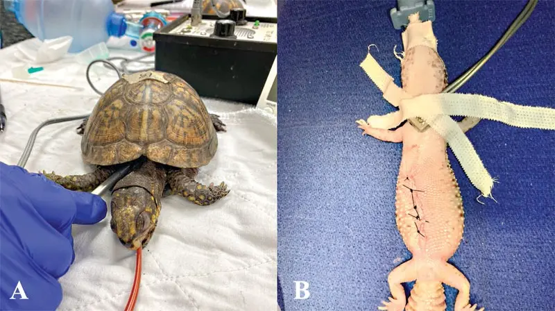
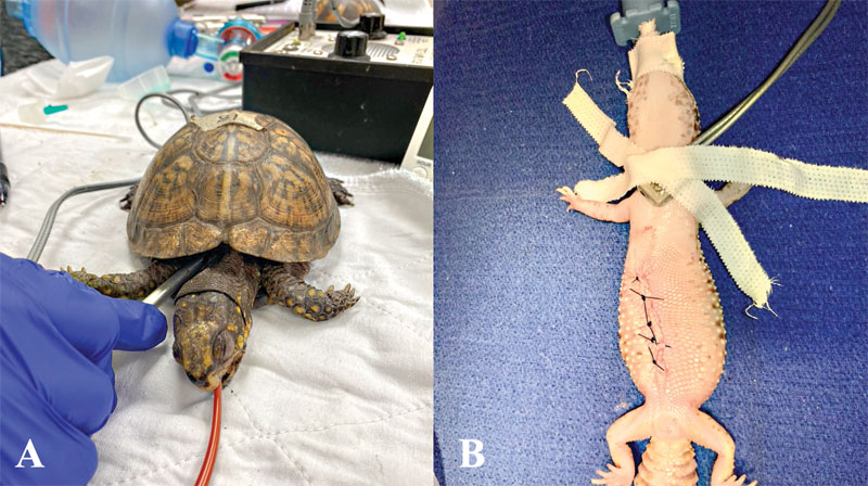
Reptilian euthanasia presents a unique set of clinical obstacles: increased resistance to hypoxia, difficult vascular access, and exceptionally slow metabolic rates. In certain species, the euthanasia process can span hours if not managed with precise pharmacological intervention. Furthermore, as seen in fish, a reptile’s heart can continue to beat autonomously even after being surgically removed and placed in formalin.
Because electroencephalograms (EEGs) are rarely available in clinical reptile practice—and standardized normal EEG values remain unpublished for virtually all reptile species—clinicians must rely on Doppler or standard ultrasound to confirm cardiac arrest. Stethoscopes are largely ineffective due to thick, scaled skin and the bony carapace and plastron of chelonians. Clinicians should listen via Doppler for at least one to two minutes, accounting for the characteristically slow heart rates of heavily sedated reptiles.
Injectable agents (intravenous preferred, or intracardiac if the animal is entirely unresponsive) and intracoelomic routes are generally accepted by the AVMA. While inhalant anesthetics are acceptable with conditions for certain species (such as snakes and select lizards), many reptiles are capable of prolonged breath-holding and intracardiac shunting. Their extreme hypoxemia tolerance severely undermines the efficacy of inhalant anesthetics as a sole method of euthanasia.
Recent pharmacological investigations into reptile euthanasia have yielded critical safety data. A study evaluating adult leopard geckos pre-medicated with alfaxalone (20 mg/kg IM) tested several secondary euthanasia routes: intracoelomic pentobarbital (400 and 800 mg/kg), intracardiac pentobarbital (800 mg/kg), intracardiac potassium chloride (KCl at 10 mEq/kg), and intracardiac lidocaine (100 mg/kg). While intracardiac pentobarbital achieved instantaneous cardiac arrest, intracoelomic administration required 30 to 35 minutes—a clinically significant delay accompanied by localized erythema and intermittent arched postures.
Most concerningly, intracardiac KCl induced severe muscle spasms so intense that animals could right themselves post-cardiac arrest. Even more unexpected, six out of nine geckos administered intracardiac lidocaine experienced a spontaneous return of respiration and heartbeat five to six hours after initial cardiac arrest. Therefore, intracardiac lidocaine and KCl are strongly discouraged at these dosages in leopard geckos.
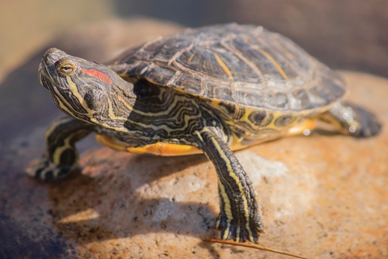
Alternative administration routes, such as oral and cloacal transmucosal delivery, have shown variable and often unreliable results. In red-eared sliders, cloacal administration of pentobarbital (100 mg/kg) resulted in significant solution leakage in 75% of subjects, necessitating rescue intravenous injections. Cardiac arrest in these animals took an average of 18 hours. While oral transmucosal delivery was more successful, it still resulted in prolonged cardiac arrest. Similarly, evaluations in bearded dragons and feral green iguanas using transmucosal pentobarbital and phenytoin demonstrated solution leakage, potential pain-related behaviors, lengthy times to cardiac arrest (averaging six hours), and a frequent need for rescue intravenous dosing.
3. Birds: Ensuring Unconscious Administration
The AVMA avian euthanasia guidelines cover a broad spectrum of birds, including companion pets, aviary species, raptors, racing pigeons, research subjects, and zoo animals (with poultry governed by separate provisions). Acceptable methods for non-poultry birds include intravenous (IV) injection of a barbiturate, provided the bird is unconscious.
Crucially, the AVMA explicitly states that an IV injection administered to a conscious bird can induce fear and distress. Consequently, deep sedation or general anesthesia is mandatory prior to injecting the euthanasia solution. The primary vessels utilized in avian patients include the right jugular vein, medial metatarsal vein, and ulnar vein. In smaller passerines and psittacines, the jugular vein is frequently the only vessel large enough to access; however, catheter placement is exceptionally challenging and typically requires heavy sedation, meaning injections are usually performed directly into the vessel.
If intravenous access is unattainable, intracardiac, intracoelomic, or intraosseous routes can be considered, provided the clinician strictly avoids air sacs and pneumatized bones (such as the humerus and femur). These alternative routes are only acceptable if the patient is fully anesthetized or unconscious. Thoracic compression in a conscious bird is strictly classified as unacceptable.
4. Small Mammals: Bridging Domestic and Exotic Protocols
Small mammals—including ferrets, rabbits, and rodents—share many physiological similarities with dogs and cats regarding euthanasia. Intraperitoneal injections are approved by the AVMA for many rodent species, though clinicians should note that time to cardiac arrest can be notably delayed compared to intravenous or intracardiac routes.
Because rabbits and rodents are prey species prone to acute stress, heavy sedation or general anesthesia is strongly recommended prior to administering euthanasia solutions. When intravenous access is complicated by hemodynamic compromise, intracardiac injection in an unconscious patient serves as a viable alternative. Additionally, an intrarenal route for rabbit euthanasia has been described and approved by the AVMA for unconscious animals; however, studies indicate that intravenous administration is significantly faster, with 30% of intrarenal subjects requiring rescue IV pentobarbital doses.
Supporting Context & Protocols
To assist veterinary practitioners in designing humane, species-appropriate protocols, the following guidelines offer structured frameworks for clinical application. Note: Dosages and procedures serve as clinical examples and must be tailored to individual patient health status and institutional protocols.
Example Fish Euthanasia Protocol
- Anesthetic Induction: Immerse the fish in buffered MS-222 (300–500+ mg/L) for a minimum of 30 minutes following the cessation of opercular movement.
- Secondary Method:
- Pithing or decapitation, OR
- Intracardiac injection of potassium chloride (KCl at 10 mEq/kg) or pentobarbital.
Example Reptile Euthanasia Protocol
- General Anesthesia Induction:
- Alfaxalone (15–20 mg/kg IM in the forelimbs), OR
- Dexmedetomidine (0.1 mg/kg IM) ± ketamine (5–20 mg/kg IM in the forelimbs), OR
- Propofol (10–20 mg/kg IV via caudal tail vein).
- Vascular Access: Intravenous or intracardiac injection of a barbiturate euthanasia solution.
- Secondary Confirmation: Additional intravenous or intracardiac KCl administered once deep unconsciousness is verified.
Example Pet Bird Euthanasia Protocol
- Heavy Sedation: Butorphanol (2–5 mg/kg IM) combined with midazolam (2–5 mg/kg IM) ± ketamine (10–20 mg/kg IM).
- Anesthetic Maintenance: Induction of general anesthesia via face mask using an inhalant anesthetic (e.g., isoflurane or sevoflurane).
- Euthanasia Delivery: Intravenous injection of a barbiturate solution via the right jugular, medial metatarsal, or ulnar vein.
Example Small Mammal (Rabbit/Rodent) Euthanasia Protocol
- Heavy Sedation: Butorphanol (2 mg/kg IM), midazolam (2 mg/kg IM), and ketamine (15–20 mg/kg IM).
- Anesthetic Maintenance: General anesthesia via mask with an inhalant anesthetic.
- Final Administration: Intravenous or intracardiac injection of a barbiturate euthanasia solution.
Environmental and Legal Considerations: Disposal of Remains
The responsibilities of the veterinary team do not end with the cessation of heartbeat and brain function. Proper disposal of animal remains is both a biosecurity necessity and an environmental safety mandate.
While communal or individual cremation/incineration is the clinical ideal, owners frequently request the return of remains for domestic burial. Veterinarians must counsel clients rigorously regarding the environmental risks associated with barbiturate retention. Pentobarbital does not break down rapidly in tissue and poses a severe risk of secondary intoxication—and frequently fatal poisoning—to wildlife scavengers, including raptors such as bald eagles. Furthermore, municipal and regional laws regarding carcass burial vary widely. Veterinary practices should mandate signed euthanasia consent forms that explicitly educate clients on proper disposal methods and secondary toxicity risks.
Future Outlook
As exotic animal medicine continues to evolve as a recognized specialty, veterinary research must address lingering gaps in euthanasia science. Future initiatives should focus on establishing normative electroencephalographic (EEG) data across a wider array of reptilian and avian species, refining non-painful transmucosal delivery systems that eliminate solution leakage, and formulating reliable, rapid protocols for hypersensitive aquatic and amphibious taxa. By anchoring clinical practice in rigorous scientific inquiry and adherence to updated AVMA frameworks, veterinary professionals can ensure that the final act of care delivered to exotic companions is defined by absolute dignity, safety, and unwavering compassion.