
By Veterinary News Desk
Special Clinical Report
Executive Overview
When a mature feline patient presents with clinical signs that include blood-tinged ptyalism (bloody drool), progressive anorexia, and severe dysphagia (difficulty swallowing), the diagnostic needle almost invariably points toward one primary suspect: sublingual or lingual squamous cell carcinoma (SCC). Statistically, playing the odds in a 10-year-old cat with these hallmark symptoms makes SCC the most probable diagnosis. However, relying solely on statistical probability in veterinary medicine can introduce critical diagnostic blind spots.
Recent clinical insights shared by Dr. John R. Lewis, VMD, DAVDC, Fellow-AVDC OMFS, highlight the imperative for veterinary practitioners to broaden their clinical horizons. Sublingual abnormalities in cats are not monospecific; a diverse array of non-neoplastic, inflammatory, and traumatic conditions can thoroughly mimic the macroscopic appearance of aggressive malignancies. From sialoceles and autoimmune-driven inflammatory dermatoses to hidden foreign bodies and chronic mechanical irritation, clinicians must systematically evaluate a wider spectrum of rule-outs before arriving at a definitive diagnosis.
This comprehensive review explores the less common, yet clinically critical, sublingual lesions that every small animal practitioner should maintain on their differential diagnosis list. By examining ranulas, eosinophilic granuloma complexes, pseudoranulas, and linear foreign bodies, this article provides an authoritative roadmap to navigating feline sublingual pathology with precision, emphasizing the irreplaceable value of histopathology and targeted interdisciplinary care.
Detailed Chronology and Diagnostic Case Studies
To understand the diagnostic challenges inherent in sublingual lesions, one must trace how these conditions present, evolve, and mimic one another in clinical settings. The diagnostic journey often begins with a routine oral examination that yields unexpected visual complexities.
The Ranula: Unraveling the Frog-Belly Swelling
When a smooth, fluid-filled swelling develops beneath a cat’s tongue, the primary consideration should be a ranula—a term derived from the Latin word rana, meaning "frog," due to its striking resemblance to a frog’s underbelly. Ranulas are fundamentally linked to the abnormal extravasation and accumulation of saliva resulting from trauma, obstruction, or rupture of salivary ducts or glands.
To fully grasp the anatomy of a ranula, clinicians must review the sublingual floor. If one lifts the tongue of a feline or canine patient, paired raised structures near the midline of the caudal mandibular symphysis become visible. These are the caruncles, which serve as the primary duct openings for the mandibular and sublingual salivary ducts. When structural damage compromises these caruncles or the delicate ducts running between the glandular tissue and their oral openings, saliva leaks into the surrounding interstitial tissues. This dynamic creates a unilateral or bilateral sublingual swelling that is typically non-inflamed and fluctuant upon palpation.
While ranulas can occasionally undergo spontaneous resolution without medical or surgical intervention, they frequently persist and enlarge. As they expand, they become vulnerable to secondary trauma from opposing dentition, compounding the patient’s discomfort. Definitive management often requires surgical intervention, specifically the dissection and excision (sialoadenectomy) of the mandibular and sublingual salivary glands.
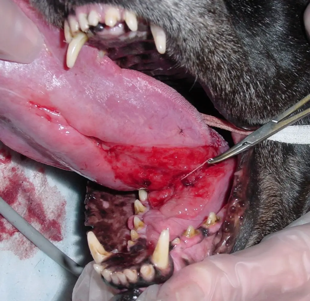
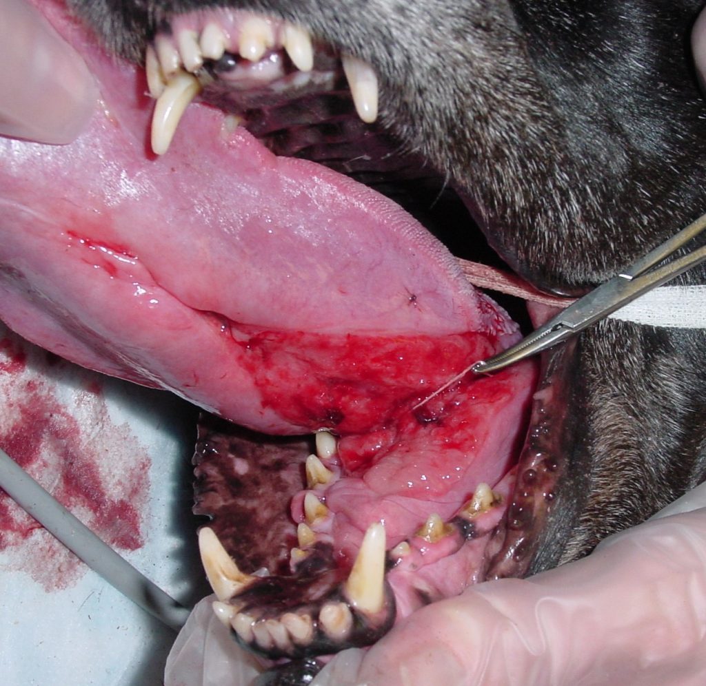
A critical diagnostic pitfall arises in the wake of oncologic surgeries. When veterinary dental surgeons perform mandibulectomies for oral tumors, transecting and ligating the salivary ducts is sometimes unavoidable. In these instances, postoperative sublingual edema and swelling can closely mimic a true ranula. However, these iatrogenic "pseudoranulas" are self-limiting and typically resolve spontaneously within a few days as the postoperative edema subsides.
Interestingly, historical veterinary literature introduces conflicting data regarding the pathogenesis of these lesions. An experimental study published in 1978 demonstrated that surgically induced trauma to salivary ducts and glands does not invariably result in abnormal salivary accumulation; in that small sample cohort, no ranulas developed following deliberate surgical trauma. Despite this experimental anomaly, contemporary clinical reality confirms that ranulas can—and do—occur postoperatively following ductal transection during oncologic procedures, reinforcing the need for vigilant postsurgical monitoring.
Eosinophilic Granulomas: The Great Mimics
While the classic textbook presentation of the feline eosinophilic granuloma complex involves ulceration of the upper lip—frequently termed "rodent ulcers"—feline eosinophilic disease exhibits a remarkable polymorphism. Non-ulcerative manifestations, including localized tissue proliferation and swelling of the lips or sublingual spaces, must be kept high on the differential diagnosis list.
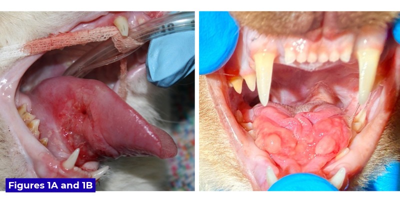
Sublingual eosinophilic granulomas are notorious diagnostic chameleons, capable of mimicking both squamous cell carcinomas and ranulas. In some instances, an eosinophilic sublingual lesion presents with a deceptively smooth surface, cleanly masquerading as a ranula. More frequently, however, they present diagnostic puzzles that closely mirror aggressive oral neoplasms.
A prime illustration of this diagnostic trap is documented in a recent clinical case managed by Dr. Lewis alongside Dr. Kirk Herrmann, DVM, DAVDC, and Dr. Alex Juriga, DVM. The patient, Milo, a one-year-old Ragdoll cat, was presented for evaluation of a significant sublingual swelling that immediately raised alarms for squamous cell carcinoma. Clinically, the sublingual mass was verrucous (wart-like), ulcerated, and remarkably firm to the touch. Compounding the clinical picture, Milo possessed a documented history of a swollen lower lip, which remained visibly enlarged at the time of presentation.
Faced with such an aggressive macroscopic appearance, the clinical team initiated an immediate diagnostic workup. Because the severity of the sublingual swelling placed Milo at high risk for ongoing self-trauma from his own teeth, medical management was initiated concurrently with diagnostic testing. Histopathologic evaluation of an incisional biopsy revealed marked eosinophilic granulomatous glossitis with concurrent ulceration—a definitive diagnosis of an eosinophilic granuloma.
Pending histopathology confirmation, clinicians administered dexamethasone sodium phosphate, transitioning the patient the following day to a daily oral prednisolone protocol at 1mg/kg/day. The therapeutic response was both rapid and dramatic: within one week, the sublingual swelling decreased significantly, achieving near-complete resolution after two weeks of targeted corticosteroid therapy. Furthermore, the clinical team recommended a comprehensive dermatological consultation to identify and isolate potential underlying allergic triggers driving Milo’s systemic eosinophilic condition. This case serves as a powerful reminder that visual inspection alone is insufficient; histopathology remains the gold standard for securing an accurate diagnosis.
Rarities: Linear Foreign Bodies and Chronic Irritation
Beyond neoplasia, sialoceles, and immune-mediated granulomas, clinicians must remain alert to unusual physical insults. Linear foreign bodies that wrap tightly around the lingual frenulum are well-documented hazards in veterinary medicine. Capable of causing severe sublingual edema, marked erythema, and profound swelling of the tongue body itself, linear foreign bodies are most frequently diagnosed in young, exploratory felines, though they are by no means exclusive to cats.
Dr. Lewis highlights an atypical canine case involving a 10-year-old dog named Lily. Lily presented with sublingual pathology after chewing on the inner twine of a golf ball, which subsequently became completely wrapped around the base of the tongue at the frenulum. This unusual presentation underscores that foreign body entrapment must be ruled out across species lines and age groups.
In addition to acute foreign body entrapment, chronic mechanical irritation can precipitate sublingual lesions. Prolonged contact with sharp or roughened dental surfaces—such as those created by advanced feline tooth resorption—can stimulate localized tissue proliferation, resulting in extensive sublingual granulation tissue that mimics neoplastic masses.
Supporting Context & Metrics
To contextualize the diagnostic challenges associated with feline sublingual lesions, veterinary professionals must evaluate the epidemiological and pathological data governing oral diseases in domestic cats.
- Prevalence of Oral Neoplasia: Squamous cell carcinoma accounts for roughly 60% to 70% of all malignant oral tumors in felines, making it the predominant neoplasm of the oral cavity. Its typical demographic involves geriatric cats (median age of approximately 10 to 12 years).
- Diagnostic Accuracy via Biopsy: Visual and physical examinations alone possess low specificity for differentiating between feline SCC, eosinophilic granulomas, and infected ranulas. Studies indicate that incisional biopsy paired with histopathology alters the initial clinical working diagnosis in up to 30% of atypical oral lesion presentations.
- Therapeutic Responsiveness: While SCC is notoriously refractory to conventional medical management—yielding poor long-term prognoses—inflammatory conditions such as eosinophilic granulomas demonstrate rapid responsiveness to corticosteroid therapy, with significant clinical reduction typically observed within 7 to 14 days of initiating protocols like prednisolone at 1mg/kg/day.
- Age Distribution Disparities: While malignant neoplasms like SCC overwhelmingly target older feline populations, inflammatory conditions (e.g., eosinophilic granulomas) and traumatic insults (e.g., linear foreign bodies) frequently present in much younger cohorts, such as one-year-old purebreds, emphasizing that patient age alone should not rule out inflammatory or traumatic etiologies.
Official Statements and Expert Perspectives
The diagnostic complexities surrounding feline sublingual lesions have drawn commentary from leading specialists in veterinary dentistry and oral surgery. The consensus among board-certified practitioners centers on moving away from presumptive diagnoses based solely on signalment and gross appearance.
"It is important to remember not every sublingual lesion in a cat is due to squamous cell carcinoma," states Dr. John R. Lewis. "Sure, if you’re playing the odds in a 10-year-old cat that presents with bloody drool, decreased appetite, and difficulty swallowing, sublingual or lingual SCC is the most likely diagnosis. However, clinical anomalies regularly challenge those odds. Keeping a broad differential diagnosis list ensures that reversible, non-neoplastic conditions are not prematurely written off as terminal malignancies."
Emphasizing the diagnostic roadmap utilized in cases like Milo the Ragdoll, Dr. Lewis and his surgical colleagues—including Dr. Kirk Herrmann and Dr. Alex Juriga—stress that biopsy is non-negotiable when dealing with atypical or aggressive-appearing oral masses.
"When an oral lesion presents as verrucous, ulcerated, and firm, the immediate psychological pull toward a diagnosis of carcinoma is strong," Dr. Lewis notes. "Yet, histopathology remains the ultimate arbiter. Without an incisional biopsy, a patient suffering from a highly treatable eosinophilic granuloma risks inappropriate prognostic counseling or premature euthanasia. Definitive diagnostics protect our patients and guide our therapeutics."
Furthermore, surgical specialists underscore the importance of surgical precision and anatomical awareness when managing ranulas and navigating oncologic resections. Recognizing the distinct pathophysiology of iatrogenic pseudoranulas prevents unnecessary secondary interventions, allowing clinicians to distinguish between true salivary duct rupture and postoperative inflammatory edema.
Future Outlook
As veterinary dentistry and oral surgery continue to evolve as specialized disciplines, the management of feline sublingual pathology is poised for refinement. Several key developments will shape clinical practice in the coming years:
1. Advanced Diagnostic Imaging and Biomarkers
While histopathology remains the gold standard for differentiating between neoplastic, inflammatory, and traumatic sublingual lesions, future clinical protocols will likely incorporate advanced non-invasive imaging modalities. High-resolution ultrasound and contrast-enhanced computed tomography (CT) are expected to play increasingly prominent roles in delineating the tissue architecture of sublingual swellings, helping clinicians differentiate fluid-filled ranulas from solid carcinomas or granulomatous proliferations prior to surgical biopsy. Furthermore, the identification of salivary and serum inflammatory biomarkers may soon assist in rapidly screening for immune-mediated conditions like the eosinophilic granuloma complex.
2. Interdisciplinary Collaboration in Feline Dermatology and Dentistry
The intersection of oral pathology and systemic dermatologic disease—as demonstrated in cases of feline eosinophilic granuloma complex—highlights the necessity for a unified, interdisciplinary approach to patient care. Future therapeutic paradigms will increasingly bridge veterinary dentistry and veterinary dermatology. By combining targeted oral surgery or localized medical management with comprehensive dermatological allergy testing and hyposensitization protocols, clinicians can address the root immunological causes of recurrent sublingual lesions, thereby reducing reliance on long-term systemic corticosteroid therapy.
3. Enhanced Educational Outreach on Diagnostic Rigor
As veterinary literature continues to spotlight atypical clinical presentations, veterinary training programs and continuing education platforms will place greater emphasis on avoiding diagnostic bias. Teaching clinicians to resist the temptation of relying purely on statistical probabilities in geriatric feline patients will reduce diagnostic errors. By integrating mandatory biopsy protocols into standard clinical workflows for all atypical sublingual lesions, the veterinary community will ensure higher standards of patient care, improved prognostic accuracy, and optimal therapeutic outcomes for feline patients facing complex oral disease.