
Executive Overview
Acute abdominal crises in pediatric and young adult dogs demand immediate, systematic diagnostic intervention and often surgical exploration. In small animal emergency practice, presentations involving persistent vomiting, progressive lethargy, and overt abdominal discomfort in a patient under one year of age immediately heighten the clinical index of suspicion for mechanical gastrointestinal obstruction.
This investigative case study details the clinical presentation, diagnostic pathway, surgical management, and underlying pathophysiology of an 11-month-old German Shepherd Dog presenting with a 48-hour history of severe acute gastrointestinal signs. Authored and managed under the clinical insights of Kendra Freeman, DVM, MS, DACVS (Large Animal/Small Animal), with diagnostic imaging contributed by Dr. Amanda Rouse, this case serves as a quintessential platform for examining one of the more devastating abdominal emergencies encountered in small animal surgery: intussusception.
Intussusception occurs when a segment of the gastrointestinal tract (the intussusceptum) telescopes into the lumen of an immediately adjacent segment (the intussuscipiens). This pathological telescoping initiates a cascade of vascular compromise, mechanical obstruction, wall edema, and, if left uncorrected, full-thickness necrosis, bowel perforation, and septic peritonitis.
By analyzing this case through the framework of veterinary surgical best practices, epidemiological realities, and current literature, this report unpacks the diagnostic dilemmas, anatomical predilections, etiological categorizations, and surgical prophylactic techniques required to manage canine intussusception successfully. Furthermore, this article explores the broader context of emergency gastrointestinal surgery, offering a deep-dive educational resource for veterinary practitioners, technicians, and students striving for clinical excellence.
Detailed Chronology: From Initial Presentation to Definitive Intervention
The 48-Hour Window of Decline
The patient in this case is an 11-month-old intact male German Shepherd Dog—an adolescent large-breed canine. The owner reported a strict 48-hour history of acute-onset, non-productive to bilious vomiting, profound lethargy, and visible behavioral signs of abdominal discomfort (including persistent stretching, reluctance to lie down, and a hunched posture).
According to the initial triage log, the patient presented with mild tachycardia, tacky mucous membranes indicative of moderate dehydration, and a tense, painful cranial and mid-abdominal palpation. No palpable sausage-shaped abdominal mass was definitively isolated on initial superficial palpation due to the patient’s tense abdominal wall guarding and moderate overall body condition, though a vague tubular structure was suspected in the right mid-abdomen.
In pediatric and young adult large-breed dogs, a 48-hour history of protracted vomiting accompanied by lethargy and fluid sequestration within the gastrointestinal tract rapidly precipitates hypovolemic shock, electrolyte derangements (specifically hyponatremia, hypokalemia, and hypochloremic metabolic alkalosis resulting from upper gastrointestinal fluid losses), and acute kidney injury secondary to pre-renal azotemia.
Diagnostic Workup and Imaging
Immediate intravenous catheterization and aggressive isotonic crystalloid fluid resuscitation were initiated to stabilize the patient’s hemodynamic parameters prior to definitive diagnostic imaging. A complete blood count (CBC) and serum biochemistry profile revealed a stress leukogram characterized by a mature neutrophilia with a mild lymphopenia, alongside elevated blood urea nitrogen (BUN) and creatinine values consistent with dehydration.
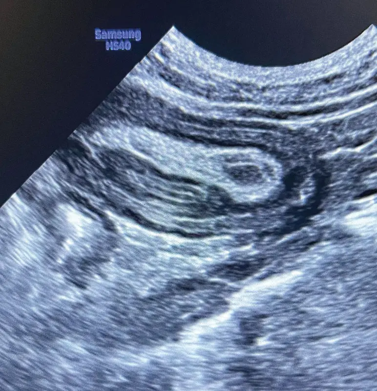
To confirm the suspected mechanical obstruction, diagnostic abdominal ultrasonography was performed by Dr. Amanda Rouse. Ultrasonography remains the gold standard imaging modality for diagnosing canine intussusception, vastly outperforming plain abdominal radiography due to its superior capability in evaluating bowel wall layering, motility, and vascular perfusion.
The ultrasonographic evaluation revealed the classic pathognomonic sign of an intussusception: the "bull’s-eye" or "target" sign when viewed in cross-section, and the "hayfork" or "layered" sign when visualized in longitudinal section. The scan demonstrated concentric rings of bowel wall within the lumen of the receiving segment. Specifically, the ultrasound
confirmed that a segment of the distal small intestine had telescoped into the colon, indicating an ileocolic intussusception. Furthermore, ultrasonography allowed the clinician to assess the viability of the trapped bowel: loss of normal wall stratification, hypoechoic thickening of the bowel wall, and absent Doppler flow within the intussusceptum signaled transmural ischemic compromise, dictating the necessity of immediate surgical exploration.
Surgical Intervention
The patient was premedicated, induced, and maintained under general anesthesia for exploratory laparotomy. Upon entering the abdominal cavity via a ventral midline celiotomy, an extensive ileocolic intussusception was identified. The terminal ileum and cecum had severely telescoped through the ileocolic junction into the ascending colon.
Due to the chronicity (48 hours) and severity of the telescoping, the affected tissues exhibited profound serosal congestion, edema, and localized fibrin deposition. Gentle manual reduction (taxis)—attempted by pushing rather than pulling the tissues apart—revealed that a significant portion of the invaginated ileum and cecum had sustained irreversible ischemic necrosis. Consequently, a conservative manual reduction alone was insufficient and contraindicated due to the high risk of subsequent perforation and peritonitis.
The surgical team performed an en bloc resection of the necrotic segments, including the terminal ileum, cecum, and proximal ascending colon, followed by a primary functional end-to-end or side-to-side anastomosis (specifically an ileocolic anastomosis) to restore intestinal continuity. The abdomen was thoroughly lavaged with warm sterile saline, and the surgical site was closed routinely. Postoperative pain management consisted of a multimodal approach incorporating constant-rate infusions (CRIs) of opioids, ketamine, and local locoregional nerve blocks, alongside supportive nutritional management initiated via early enteral feeding.
Supporting Context & Metrics: Unpacking the Brainteaser Challenges
To further contextualize the clinical decision-making involved in this case, we examine the fundamental veterinary questions presented in the clinical challenge:
+-----------------------------------------------------------------------------+
|                     CANINE INTUSSUSCEPTION: QUICK FACTS                     |
+-----------------------------------------------------------------------------+
| Most Common Anatomical Location : Ileocolic Junction                        |
| Primary Predisposing Etiology   : Idiopathic / Parasitic (in puppies)       |
| Surgical Recurrence Risk        : Mitigated via proper resection/plication  |
+-----------------------------------------------------------------------------+
Challenge Question 1: Anatomical Predilection
In dogs, the most common location for an intussusception is:
a. Ileocolic junction
b. Jejunojejunal
c. Jejunoileal
d. Coloncolic
Correct Answer: a. Ileocolic junction
In-Depth Analysis:
Intussusceptions can theoretically occur at any point along the gastrointestinal tract, but epidemiological studies consistently demonstrate that the ileocolic junction is overwhelmingly the most frequent site in dogs, accounting for up to 40% to 70% of all reported cases.
The high incidence at this specific anatomical transition zone is primarily driven by physiological and anatomical factors. The terminal ileum is relatively fixed in its position relative to the more mobile cecum and ascending colon. This abrupt fixation point, combined with the distinct peristaltic contractions of the small intestine meeting the large capacity of the ascending colon, creates a biomechanical vulnerability. When hypermotility occurs—often triggered by enteritis, parasitism, or dietary indiscretion—the terminal ileum is easily propelled or sucked into the wider lumen of the colon.
While jejunojejunal and jejunoileal intussusceptions do occur (particularly secondary to linear foreign bodies or localized mass lesions), their overall clinical frequency is significantly lower than that of the ileocolic variety. Coloncolic intussusceptions are exceedingly rare in canine patients.
Challenge Question 2: Etiological Landscape
The most common underlying cause of canine intussusceptions is:
a. Parasitic
b. A foreign material
c. Neoplasia
d. Idiopathic
Correct Answer: d. Idiopathic (Though heavily intertwined with parasitic enteritis in young cohorts)
In-Depth Analysis:
The etiology of canine intussusception is multifactorial, but it is heavily age-dependent. When evaluating pediatric populations (dogs under one year of age, such as this 11-month-old German Shepherd Dog), the vast majority of cases are classified as idiopathic, meaning no specific structural or neoplastic lesion is identified as the primary trigger. However, underlying functional dysmotility is invariably present.
Common contributory factors in young animals include:
- Parasitic Infestations: Heavy burdens of endoparasites (such as Parascaris, Ancylostoma, or Trichuris species) induce severe local enteritis, mucosal irritation, and chaotic, uncoordinated peristaltic waves that predispose the bowel to telescope.
- Infectious Enteritis: Viral pathogens (e.g., Canine Parvovirus) cause profound mucosal destruction, sloughing, and dysmotility, frequently resulting in intussusceptions.
- Dietary Indiscretion: Abrupt diet changes, ingestion of indigestible roughage, or foreign materials can alter normal propulsive motility.
Conversely, in mature and geriatric dogs, intussusceptions are much more frequently secondary to structural abnormalities acting as a "lead point." These include intestinal neoplasia (such as adenocarcinoma, lymphosarcoma, or leiomyoma), linear or focal foreign bodies, post-surgical adhesions, or severe localized granulomatous disease.
Challenge Question 3: Surgical Prophylaxis and Enteral Plication
According to Larose et al., enteral plication after manual reduction of an intussusception prevents recurrence of the intussusception.
a. True
b. False
Correct Answer: b. False
In-Depth Analysis:
Intestinal plication (or mesenteric pleating, commonly associated with the Noble plication procedure) involves suturing adjacent loops of the small intestine together in a series of gentle folds to prevent the bowel from twisting, telescoping, or re-forming an intussusception. Historically, plication was advocated by some surgeons as an aggressive prophylactic measure—particularly in recurrent cases or following simple manual reduction (taxis) where bowel viability was preserved without resection.
However, contemporary surgical literature, including clinical investigations referenced in veterinary surgical discourse (such as studies by Larose et al. and broader retrospective analyses on canine intussusception recurrence), challenges the blanket efficacy of this technique.
Critical findings regarding enteral plication include:
- Complication Risks: Plication is associated with significant morbidity, including severe intra-abdominal adhesion formation, mechanical obstruction secondary to sharp angulation of the bowel loops, inadvertent puncture of the mesenteric vessels or intestinal lumen during suturing, and localized peritonitis.
- Efficacy Deficits: Retrospective evaluations demonstrate that when an intussusception is properly managed via resection and anastomosis (when viability is compromised) or when the underlying primary cause (e.g., parasite clearance, mass removal) is successfully treated, the native recurrence rate is remarkably low, rendering aggressive prophylactic plication largely unnecessary.
- Current Consensus: Enteral plication is no longer routinely recommended as a standard prophylactic adjunct for all intussusceptions. Its use is strictly reserved for highly select, recurrent cases of idiopathic small intestinal intussusception where no resectable lead point exists, and even then, the risks versus benefits must be meticulously weighed.
Official Statements and Veterinary Expert Insights
To contextualize the management of pediatric gastrointestinal emergencies within modern veterinary medicine, veterinary surgeons emphasize a standardized, evidence-based approach to patient care.
Dr. Kendra Freeman, a board-certified large and small animal surgeon, underscores the critical nature of early surgical intervention in cases presenting with systemic compromise:
"When managing an adolescent large-breed dog with a 48-hour history of protracted vomiting and suspected intussusception, time is quite literally tissue. The window between reversible mucosal edema and catastrophic transmural necrosis is remarkably narrow. Ultrasonography has transformed our diagnostic velocity, allowing us to bypass prolonged, potentially harmful contrast series and move directly to theater. In cases where the bowel has undergone ischemic necrosis—as is frequently observed at the ileocolic junction following prolonged entrapment—attempting conservative manual reduction alone is a fatal error. Aggressive resection and meticulous restoration of gastrointestinal continuity are non-negotiable for survival."
Furthermore, emergency criticalists highlight the absolute necessity of rigorous perioperative fluid therapy and aggressive nutritional support. Because protein-losing enteropathy and third-space fluid sequestration characterize advanced gastrointestinal obstructions, patient stabilization must focus heavily on:
- Restoring intravascular volume and correcting electrolyte imbalances prior to anesthetic induction.
- Minimizing perioperative hypotension through tailored, goal-directed fluid protocols and advanced cardiovascular monitoring.
- Initiating early enteral nutrition (via post-operative trickle feedings or post-pyloric feeding tubes when indicated) within 12 to 24 hours of surgery to support enterocyte health, accelerate mucosal healing, and prevent bacterial translocation from the gut lumen.
Future Outlook: Advancements in Pediatric Gastrointestinal Surgery
The prognosis for canine patients undergoing surgical management for intussusception has improved significantly over the past two decades, largely driven by advancements in diagnostic imaging, safer anesthetic protocols, and a deeper understanding of perioperative critical care.
Looking forward, several key areas in veterinary surgery and emergency medicine are poised to further optimize outcomes for patients presenting with conditions like intussusception:
1. Enhanced Diagnostic Modalities and Point-of-Care Ultrasound (POCUS)
The widespread adoption of Point-of-Care Ultrasound (POCUS) in emergency triage settings means that primary care veterinarians and emergency clinicians can identify the characteristic "target" sign of an intussusception within minutes of presentation. Future advancements in portable, high-resolution ultrasound units integrated with artificial intelligence (AI) assistance will likely streamline pattern recognition, enabling even earlier detection before severe systemic sepsis and tissue necrosis manifest.
2. Minimally Invasive Surgical Innovations
While exploratory celiotomy remains the gold standard for resecting compromised bowel segments in emergency settings, laparoscopic-assisted and thoracoscopic techniques are rapidly evolving in veterinary surgery. For non-necrotic, reducible small intestinal intussusceptions, laparoscopic visualization and reduction techniques offer the potential benefits of reduced postoperative pain, smaller surgical incisions, and accelerated return to normal function. However, safety and speed remain paramount in acute abdomen cases; open celiotomy will remain the primary approach when bowel resection and complex anastomosis are required.
3. Regenerative Medicine and Anastomotic Healing
Research into advanced biomaterials, biologically active sealants, and regenerative medicine holds profound promise for gastrointestinal surgery. The prevention of anastomotic dehiscence—one of the most feared and lethal complications following intestinal resection—remains a major focus of clinical research. The future integration of bio-engineered serosal patches and growth-factor-infused suturing materials may further reduce leakage rates at the ileocolic junction, improving long-term survivability in complicated emergency surgeries.
4. Preventive Veterinary Medicine and Parasite Control
Ultimately, mitigating the incidence of pediatric intussusceptions relies heavily on robust preventive veterinary care. Education regarding comprehensive parasite control programs, strict adherence to vaccination schedules against viral enteropathogens (such as Canine Parvovirus), and strict management of dietary indiscretion in large-breed puppies will continue to serve as the primary defense against this devastating surgical emergency.
Conclusion
The presentation of an 11-month-old German Shepherd Dog with a 48-hour history of vomiting, lethargy, and abdominal discomfort serves as a classic textbook example of pediatric ileocolic intussusception. Through the coordinated efforts of emergency clinicians, diagnostic imagers, and board-certified surgeons, timely ultrasound identification coupled with aggressive fluid resuscitation and precise surgical resection of necrotic tissue averted a potentially fatal outcome.
By analyzing the anatomical vulnerabilities of the ileocolic junction, understanding the age-related etiological landscape, and debunking outdated surgical dogmas regarding routine enteral plication, veterinary professionals can continue to elevate the standard of care for acute abdominal emergencies in small animals.