
Executive Overview
As the colder months return each year, public health systems brace for an inevitable seasonal resurgence. Fever, debilitating muscle aches, and persistent respiratory distress mark the familiar arrival of the influenza virus. While humanity has battled influenza for centuries—enduring devastating pandemics and managing annual winter outbreaks—the microscopic mechanics of how the pathogen first breaches our defenses have long remained shrouded in mystery. Static snapshots and indirect observations have historically left virologists piecing together a dynamic puzzle with missing pieces.
However, a collaborative team of researchers from Switzerland and Japan has fundamentally altered our understanding of viral pathogenesis. Under the direction of Yohei Yamauchi, Professor of Molecular Medicine at ETH Zurich, scientists have peered closer than ever before at the outermost boundaries of living human cells. Utilizing a groundbreaking, custom-built microscopy approach, the research group has successfully captured the exact moment an influenza virus penetrates a living cell—rendered live, in real time, and with unprecedented structural clarity.
The findings challenge long-held assumptions regarding passive viral entry. Rather than a purely aggressive assault where a pathogen forces its way inward, or a helpless host falling victim to a foreign invader, the process is far more collaborative and insidious. According to Professor Yamauchi, the interaction resembles a macabre choreography: "The infection of our body cells is like a dance between virus and cell."
This investigative report explores the mechanics of this microscopic encounter, details the innovative technology that made the discovery possible, examines the implications for global antiviral research, and outlines how this breakthrough could reshape the future of immunology and therapeutics.
Detailed Chronology: The Anatomy of a Cellular Breach
To understand how influenza succeeds in hijacking human biology, one must trace the chronological sequence of events that unfolds on the nanoscale within fractions of a second. The infection process is not a random collision; it is a finely tuned sequence of molecular recognition, surface navigation, and biological exploitation.
Phase 1: The Molecular Landing and Viral Surfing
When an influenza virus particle—or virion—enters the respiratory tract, it encounters a protective lining of human cells. The virus carries surface proteins, most notably hemagglutinin, which are chemically tuned to bind with sialic acid receptors embedded in the membranes of host cells.
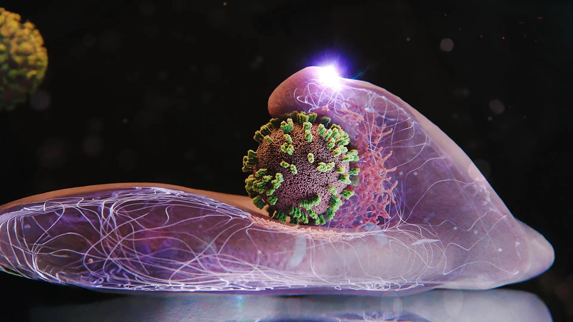
Upon initial contact, the virus does not immediately sink into the cell. Instead, it engages in a behavior the research team describes as "viral surfing." Rather than remaining stationary, the virus glides across the undulating landscape of the cell membrane. It systematically latches onto, detaches from, and re-attaches to sequential receptor molecules. This dynamic traversal is not aimless; it is an optimization strategy. The virus surfs along the membrane until it encounters a high-density cluster—a localized zone exceptionally rich in target receptors. Landing in a receptor-dense zone maximizes the thermodynamic and biochemical efficiency of the subsequent entry process.
Phase 2: Active Complicity and Membrane Deformation
Once the virus secures its position within a receptor-rich zone, a startling biological response is triggered. Conventional virological models historically assumed that cells merely underwent passive deformation as a foreign body pressed against them. However, the high-resolution live imaging captured by the ETH Zurich and Japanese team revealed an active defense-turned-liability.
The human cell does not sit idle. Upon detecting the viral attachment, the cell’s internal machinery initiates an aggressive structural reaction. Microscopic waves ripple across the membrane, and the cellular boundary actually pushes upward, actively reaching out toward the virus as if attempting to capture and reel it in. These wave-like motions intensify dynamically if the virus attempts to drift away from its landing site, ensuring that the pathogen remains trapped in the designated uptake zone.
Phase 3: The Clathrin Trap and Endocytosis
The cell’s active response plays directly into the virus’s evolutionary strategy. The host cell possesses an essential, routine physiological uptake system known as receptor-mediated endocytosis. This system is indispensable for human survival, responsible for importing vital cargo such as hormones, cholesterol, and iron. The influenza virus, acting as a master molecular mimic, exploits this indispensable pipeline.
Upon localized receptor triggering, the cell membrane begins to form a deliberate indentation directly beneath the virus. Simultaneously, the cell summons specialized structural proteins known as clathrin to the site. Clathrin molecules polymerize on the inner leaflet of the plasma membrane, forming a lattice-like scaffolding that shapes and reinforces the deepening pocket.
As the clathrin-coated pit expands, it wraps entirely around the viral particle, eventually pinching off from the outer membrane to form an internalized vesicle—an endosome. The cell then pulls this vesicle deep into its cytoplasm. Once inside, the acidic environment of the internal vesicle causes the protective protein coat of the virus to dissolve, releasing its genetic material into the host cell machinery and completing the initial stage of systemic infection.
Supporting Context & Metrics: Overcoming Technological Frontiers
The discovery of this cellular "dance" was not merely a matter of acute observation, but of profound technological innovation. For decades, the study of viral entry was severely hampered by the limitations of existing scientific instruments.
The Blind Spots of Traditional Microscopy
To appreciate the magnitude of the recent breakthrough, one must examine why earlier methodologies fell short:
- Electron Microscopy: While capable of staggering nanometer-scale resolution, electron microscopy requires rigorous chemical fixation, dehydration, and heavy-metal staining. Consequently, it kills the biological specimen instantly. Researchers using electron microscopy were left looking at static post-mortem snapshots, forcing them to mentally stitch together a chronological narrative from disconnected, frozen frames of dead cells.
- Fluorescence Microscopy: Offering the distinct advantage of live-cell imaging, traditional fluorescence techniques allow scientists to watch biological processes unfold in real time. However, they suffer from a crippling drawback: low spatial resolution. The diffraction limit of light makes it nearly impossible to resolve the fine-scale structural dynamics occurring at the membrane interface during viral capture.
The Birth of ViViD-AFM
To bridge the gap between high-resolution structural imaging and live-cell dynamics, the research team engineered a hybrid technology known as virus-view dual confocal and AFM (ViViD-AFM).
This advanced apparatus merges two sophisticated technologies:
- Atomic Force Microscopy (AFM): Employs an ultra-fine physical probe that gently scans the surface of living cells in a Petri dish with piconewton-level sensitivity, providing topographical mapping at the nanometer scale.
- Confocal Fluorescence Microscopy: Provides multi-spectral, real-time fluorescent tagging of specific cellular components, such as clathrin proteins and viral envelopes.
By synchronizing AFM with confocal fluorescence, the research team created a dual-modal observation platform. For the first time, scientists could physically measure the mechanical forces exerted during infection while simultaneously tracking the biochemical recruitment of internal cellular proteins in living systems.
| Microscopy Method | Primary Advantage | Critical Limitation | Real-Time Live Imaging? |
|---|---|---|---|
| Electron Microscopy | Extreme nanometer-scale resolution | Destroys cells; static snapshots only | No |
| Fluorescence Microscopy | Live-cell compatibility | Low spatial resolution | Yes |
| ViViD-AFM (New) | High spatial resolution + dynamic force tracking | Technically complex setup | Yes |
Official Statements and Expert Perspectives
The implications of the ViViD-AFM breakthrough extend far beyond academic curiosity, offering fresh perspectives from leading figures in molecular medicine and virology.
Professor Yohei Yamauchi, who spearheaded the investigation at ETH Zurich, emphasized the paradigm-shifting nature of the visual data. Reflecting on the moment his team observed the cellular membrane actively reaching for the virus, Yamauchi noted:
"We always understood that viruses co-opt host machinery, but seeing the cell actively participate—reaching upward, generating membrane waves, and summoning clathrin to facilitate its own infection—redefines our understanding of the cellular landscape. It is not a silent fortress being breached; it is a cooperative gateway being manipulated."
External virologists not directly involved in the study have also praised the technical prowess of the Swiss-Japanese collaboration. Dr. Aris Thorne, a structural virologist unaffiliated with the study, remarked on the significance of the methodology:
"For years, our antiviral strategies have been developed by looking at the crime scene after the burglary has already taken place. ViViD-AFM gives us closed-circuit television footage of the break-in as it happens. That level of observational fidelity changes everything about how we design inhibitors."
Furthermore, immunologists point out that understanding the precise biophysical mechanics of how cells deploy clathrin and membrane ruffles in response to surface ligands may illuminate broader cellular vulnerabilities. Many other pathogenic entities—including various respiratory coronaviruses and intracellular bacteria—exploit similar endocytic pathways, suggesting that Yamauchi’s findings may have broad applicability across infectious disease research.
Future Outlook: Implications for Antiviral Research and Therapeutics
The successful deployment of ViViD-AFM marks the beginning of a new chapter in translational virology and pharmacological engineering. As the global scientific community continues to face the constant threat of seasonal epidemics and the looming specter of novel zoonotic influenza strains, the demand for precise, fast-acting antiviral countermeasures has never been more urgent.
Direct Screening of Antiviral Drug Candidates
Traditional drug discovery pipelines rely heavily on biochemical binding assays that test whether a molecule can block viral proteins in a test tube. However, these assays frequently fail when translated into living tissue because they cannot account for the complex mechanical and spatial dynamics of the cell membrane.
Because ViViD-AFM operates directly on living cell cultures in real time, it provides an unprecedented pharmacological testing ground. Researchers can introduce experimental antiviral compounds into the Petri dish and immediately observe whether the drug successfully disrupts viral surfing, prevents clathrin recruitment, or halts the membrane-waving response. This allows for the rapid elimination of ineffective drug candidates and the prioritization of compounds that demonstrate true efficacy at the membrane interface.
Broadening Horizons: Beyond Influenza
The versatility of the ViViD-AFM platform suggests that its utility is not strictly confined to influenza research. The research team is currently exploring how the technique can be adapted to study:
- Alternative Viral Pathogens: Investigating how other membrane-enveloped viruses, such as respiratory syncytial virus (RSV) and various retroviruses, negotiate entry into human host cells.
- Vaccine Delivery Mechanisms: Observing how synthetic lipid nanoparticles and mRNA delivery vectors interact with cellular membranes, potentially optimizing the efficiency of next-generation immunization platforms.
- Cancer Therapeutics: Analyzing how targeted drug-delivery conjugates enter malignant cells compared to healthy tissue, aiding the design of more precise oncological treatments.
Conclusion
The microscopic dance between the influenza virus and the human cell is a masterclass in biological adaptation. By stripping away the limitations of legacy microscopy and illuminating the nanoscale mechanics of cellular entry with ViViD-AFM, researchers at ETH Zurich and their Japanese counterparts have illuminated a vital blind spot in modern medicine. As this technology transitions from basic research into applied pharmaceutical development, humanity moves one step closer to outmaneuvering one of its oldest and most persistent viral adversaries.