
By John Lewis, VMD, DAVDC, Fellow-AVDC OMFS
Executive Overview
Veterinary dentistry has experienced a profound evolution over the past two decades, transitioning from a mechanical, force-heavy discipline to a refined surgical specialty driven by an intimate understanding of oral anatomy and specialized instrumentation. Building on last month’s exploration of fundamental diagnostic and therapeutic tools, this installment delves into the advanced equipment, procedural nuances, and anatomical mechanics that render complex tooth extractions safer, more efficient, and less traumatic for veterinary patients.
At the core of every successful extraction lies the management of the periodontium—the complex array of tissues anchoring the tooth to the alveolar bone. Safely delivering a firmly rooted tooth requires the systematic disruption of millions of microscopic periodontal ligament (PDL) fibers. Historically, veterinary practitioners relied heavily on brute force, mechanical leverage, and crude pulverization techniques that often led to protracted recovery times, iatrogenic bone fractures, and unnecessary surgical morbidity.
Today, contemporary veterinary dental practitioners utilize an array of micro-engineered instruments. From thin-profile winged elevators and delicate luxators to automated periotomes, piezosurgical units, and specialized surgical burs, modern technology prioritizes finesse over force. By understanding the biomechanical properties of the periodontium and leveraging state-of-the-art armamentarium, clinicians can dramatically reduce chair time, minimize tissue trauma, and elevate the standard of patient care across both general and specialty veterinary practices.
Detailed Chronology and Technical Evolution of Extraction Instrumentation
The progression of veterinary oral surgery mirrors developments in human dentistry, shifting from rudimentary extraction forceps to sophisticated, tissue-specific preservation tools. To appreciate modern extraction techniques, one must trace the step-by-step procedural chronology of a modern veterinary dental extraction, examining how specialized instruments are deployed at each phase of the surgical intervention.
Phase 1: Soft Tissue Incision and Flap Elevation
The extraction process invariably begins with the liberation of the gingival attachment. This initial phase ensures direct visualization of the alveolar bone and prevents tearing of the delicate gingival margin during subsequent root mobilization.
- The 15C Scalpel Blade: Widely regarded as the gold standard among veterinary dental specialists, the 15C blade features a curved cutting edge ideal for precision incising of the gingival connective tissue. It allows for clean intrasulcular incisions and precise releasing incisions when mucogingival flaps are required to expose multi-rooted teeth or compromised roots.
- Periosteal Elevators: Once the initial sulcular incision is made, sharp periosteal elevators are deployed to gently lift the mucogingival flap away from the underlying alveolar bone. The sharpness of these instruments is paramount; dull periosteal elevators tend to shred tissue rather than cleanly cleave the periosteum from the bone, thereby increasing postoperative discomfort and delaying healing.
Phase 2: Disruption of the Periodontal Ligament
With the alveolar bone exposed (when flaps are utilized) or directly accessible through the sulcus, the primary challenge of the extraction emerges: overcoming the structural integrity of the periodontium.
The periodontium is composed of the root surface (cementum), the alveolar bone, the gingival connective tissue, and the periodontal ligament. The PDL occupies the narrow space between the alveolar bone and the cementum, functioning as a shock absorber during mastication. Biochemically, the PDL is densely populated—primarily consisting of type I, III, and V collagen fiber bundles, an amorphous ground substance matrix, and a rich vascular network. Collagen fiber bundles alone account for an estimated 50 to 75 percent of the total volume of the periodontal ligament.
To successfully extract a tooth, these millions of microscopic collagen bundles must be systematically stretched, torn, or severed. Modern instrumentation approaches this task through specialized mechanisms:
- Winged (Wiggs) Dental Elevators: Characterized by thin, lateral "wings" designed to conform to the root contour, winged elevators are seated directly into the periodontal ligament space. When the instrument handle is rotated, the wings engage the root and the opposing bone, leveraging the tooth while simultaneously stretching and fracturing the PDL fibers.
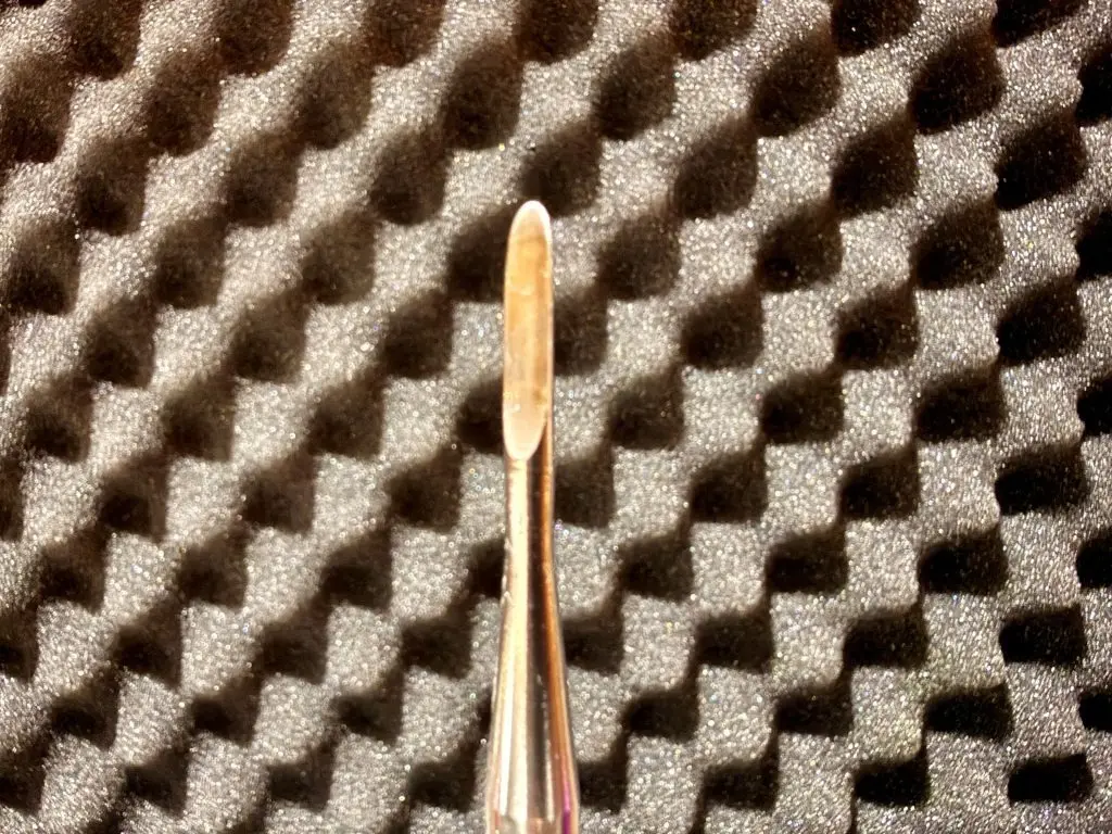
- Dental Luxators: While elevators rely partially on rotational leverage, luxators function via a cutting mechanism. Engineered like miniature scalpel blades, luxators are inserted into the PDL space and manipulated in a gentle, side-to-side windshield-wiper motion. This action extends the blade deeper into the alveolus, surgically severing the collagen fibers rather than simply tearing them through force. Because the average canine or feline periodontal ligament space is approximately 0.25 mm (250 microns) wide, luxator blades must be exceptionally thin. Consequently, they require frequent sharpening or replacement to maintain their cutting edge.
Phase 3: Advanced Mechanical and Ultrasonic Assistance
For ankylosed teeth (where the root is fused directly to the alveolar bone) or exceptionally robust, multi-rooted teeth, standard hand instruments may prove insufficient. Modern operatories increasingly incorporate power-assisted technology:
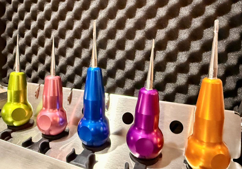
- Automated Periotomes: Functioning on a reciprocating, pneumatic principle—much like a miniature jackhammer—automated periotomes rapidly drive an ultra-thin blade into the PDL space. While exceptionally efficient in normal periodontia, they are contraindicated in cases of ankylosis due to the risk of catastrophic tip breakage.
- Piezosurgical Units: Piezosurgery has transformed oral and maxillofacial surgery. Utilizing ultrasonic micro-vibrations, piezosurgical units are routinely employed for complex procedures such as maxillectomies and mandibulectomies. In extraction settings, specialized piezosurgical tips can be advanced up to 10 mm into the periodontal space to liberate attachments, and unlike automated periotomes, they can be effectively utilized even in the presence of ankylosis.
Phase 4: The "Moat" Technique and Root Management
When dealing with fractured, retained, or severely compromised roots, traditional methods often involved "pulverizing" or "atomizing" the root with a large bur—a technique fraught with the danger of thermal necrosis, iatrogenic bone damage, and incomplete root removal.
Modern veterinary surgery favors the moat technique. This protocol utilizes a high-speed surgical handpiece equipped with a miniature, long-shafted bur (such as a ¼ surgical length round bur) to carve a precise "moat" around the periphery of the stubborn root. Simultaneously, a small window is created in the buccal alveolar bone. This process exposes the root boundary and provides a precise point of entry for a small luxator, Davis root tip elevator, or feline periosteotome. Gentle, controlled leverage via this created space delivers the root cleanly and atraumatically.
Supporting Context & Anatomical Metrics
To fully appreciate the rationale behind modern veterinary dental instrumentation, clinicians must examine the quantitative and biological parameters governing tooth extractions:
| Parameter / Metric | Clinical Specification | Surgical Significance |
|---|---|---|
| Periodontal Ligament Width | ~0.25 mm (250 microns) | Demands ultra-thin instrument profiles (e.g., specialized luxators) to enter the space without causing crushing trauma to surrounding bone. |
| PDL Composition Volume | 50% – 75% Type I, III, and V collagen | Dictates the need for sharp cutting instruments (luxators) rather than blunt force, as collagen bundles offer high tensile strength. |
| Piezosurgery Penetration Depth | Up to ~10 mm | Allows safe advancement along root surfaces, even in compromised or ankylosed roots where automated periotomes fail. |
| Surgical Round Bur Size | ¼ surgical length round bur | The optimal size for creating a precise "moat" around stubborn root tips while preserving structural buccal bone. |
The transition from brute-force extractions to micro-surgical precision has direct financial and clinical ramifications for veterinary practices. Reducing surgical time decreases anesthetic duration, which in turn mitigates patient risk. Furthermore, preserving the integrity of the alveolar socket is critical if subsequent procedures—such as bone grafting or dental implants—are considered, or simply to ensure rapid, complication-free primary-intention soft tissue healing.
Expert Insights and Clinical Perspectives
Industry leaders emphasize that the integration of advanced instrumentation is not merely a luxury, but a fundamental ethical and clinical imperative in modern companion animal practice.
"When extracting a firmly rooted tooth, we are essentially fighting millions of years of evolutionary engineering designed to keep that tooth firmly anchored in the jaw," notes Dr. John Lewis, VMD, DAVDC, Fellow-AVDC OMFS. "By shifting our mindset from ‘how hard can I twist this elevator’ to ‘how precisely can I sever these microscopic collagen attachments,’ we completely change the risk profile of the procedure. Every successful extraction is a victory over millions of individual periodontal ligament fibers."
Specialists also stress the importance of ergonomic and safety protocols. Because delicate instruments like luxators and ¼ surgical length round burs possess extremely fine working ends, they are susceptible to metal fatigue and breakage. Dr. Lewis advises practitioners to treat delicate burs as single-use items in clinical practice and to always employ safe hand positioning:
- The Safety Stop Technique: Clinicians must keep an index finger extended along the shaft of elevators and luxators. In the inevitable event that the instrument slips during application, the practitioner’s finger acts as an immediate physical stop, preventing the sharp tip from plunging into adjacent soft tissues, the palate, or the eye.
Furthermore, modular instrumentation systems—which allow clinicians to swap out worn blades from reusable handles without discarding the entire instrument—have streamlined inventory management and ensured that cutting edges remain razor-sharp across all surgical cases.
Future Outlook and Technological Horizons
As veterinary medicine continues to borrow and adapt innovations from human maxillofacial surgery and bioengineering, the future of dental extractions points toward even greater precision, real-time imaging integration, and minimally invasive protocols.
- Enhanced Ultrasonic and Laser Integration: Future developments will likely refine piezosurgical tips further, reducing handpiece dimensions to improve intraoral access, particularly in toy-breed dogs and feline patients where anatomical working spaces are exceptionally narrow.
- Advanced Biomaterials and Socket Preservation: As root removal becomes increasingly atraumatic through techniques like the moat method, the preservation of pristine alveolar sockets will open new avenues for immediate bone-grafting materials, synthetic membranes, and regenerative biologics in veterinary patients.
- Ergonomic and Smart Instrumentation: The incorporation of torque-limiting handles and tactile feedback sensors into dental elevators and luxators may soon provide clinicians with real-time digital monitoring of applied force, virtually eliminating accidental root tip fractures caused by excessive operator leverage.
Ultimately, the mastery of veterinary extractions rests at the intersection of anatomical science and technological refinement. By embracing modern luxators, piezosurgery, and refined bur techniques, veterinary professionals can ensure safer surgeries, faster recovery for patients, and a consistently elevated standard of comprehensive oral health care.