
Executive Overview
For decades, evolutionary biologists have mapped the complex architecture of the vertebrate eye, marveling at how natural selection sculpted the intricate camera-like organs found in humans, birds, fish, and mammals. Yet, a fundamental evolutionary puzzle has continuously baffled scientists: why do vertebrate eyes possess a developmental blueprint so fundamentally distinct from those of invertebrates like insects, octopuses, and squids? While cephalopods and arthropods develop their visual apparatus outward from the ectodermal tissue of the skin, the vertebrate retina emerges directly as an extension of the embryonic brain itself.
Now, groundbreaking research spearheaded by evolutionary biologists at Lund University and the University of Sussex has turned our understanding of neurobiology and sensory evolution completely on its head. According to their findings, every living vertebrate on Earth—from deep-sea fish to human beings—shares a surprisingly bizarre common ancestor that lived nearly 600 million years ago. This distant, worm-like relative possessed a single, central eye positioned prominently on top of its head, evoking the mythological terror of a microscopic cyclops.
This provocative study suggests that our evolutionary lineage took a radical detour. Long before complex swimming vertebrates populated the ancient Cambrian oceans, an early ancestor discarded its paired eyes during a sedentary, filter-feeding phase of existence. In their place, a solitary "median eye" evolved atop the creature’s head to monitor basic light cycles. Millions of years later, when our ancestors returned to an active, swimming lifestyle, the visual system was entirely rebuilt not from the discarded ancient lateral eyes, but from this singular median structure.
Crucially, this evolutionary ghost story leaves a physical trace inside every human skull today. The modern pineal gland—a tiny, pinecone-shaped endocrine structure buried deep within the center of the human brain that regulates our sleep-wake cycles through melatonin production—is the direct evolutionary descendant of that ancient, cyclopean median eye. This report explores the chronological unfolding of this discovery, the physiological and genetic mechanics behind vertebrate vision, official insights from the lead researchers, and the profound implications this holds for the future of neuroscience and evolutionary biology.
Detailed Chronology: From Ediacaran Worms to Modern Neurobiology
To comprehend how modern human vision is tethered to a prehistoric single-eyed organism, scientists must look backward across six hundred million years of geological and biological time, tracing an evolutionary odyssey defined by environmental adaptation, physiological regression, and revolutionary architectural reinvention.
Phase I: The Ediacaran Twilight and the Loss of Sight (approx. 600 Million Years Ago)
During the Ediacaran period, long before the famous "Cambrian explosion" flooded the prehistoric seas with complex predatory animals, the ancestral lineage leading to all modern vertebrates was a modest, soft-bodied, worm-like creature. Living on or just beneath the soft sediment of ancient seabeds, this organism survived through passive suspension feeding, filtering microscopic plankton and organic detritus directly out of the seawater.
Fossil evidence and comparative genetic reconstructions suggest that even earlier in its lineage, this animal likely possessed rudimentary paired eyes—simple light-sensitive spots or rudimentary image-forming organs shared with other primitive branches of the animal tree of life. However, life as a stationary filter-feeder imposed entirely different evolutionary pressures. Moving through a complex three-dimensional environment in search of prey or fleeing from agile predators was no longer a primary daily concern.
Maintaining complex, paired lateral eyes required significant metabolic energy and genetic maintenance. Because the stationary organism spent the vast majority of its life anchored in one spot, natural selection favored economy over sensory acuity. Over countless generations, the creature’s paired lateral eyes gradually atrophied and disappeared altogether.
Phase II: The Rise of the Median Cyclops
Although the worm-like organism jettisoned its lateral eyes, total blindness in a fluctuating photic environment proved disadvantageous. Even stationary filter-feeders benefit profoundly from detecting the diurnal cycle—knowing when the sun rises and sets allows them to synchronize feeding behaviors, metabolic rates, and protective responses.
To fulfill this need, a cluster of primitive, light-sensitive cells persisted in the exact center of the creature’s head. Over millions of years of gradual refinement, these centralized cells consolidated, forming a single, primitive "median eye." Positioned on the dorsal surface of the head, this cyclopean structure was not capable of resolving sharp images of predators or prey. Instead, it operated as an environmental light sensor, capable of distinguishing between harsh daylight and deep shadow, while assisting the organism with spatial orientation relative to the water’s surface.
For an extensive epoch in early vertebrate history, this single, median eye stood as the sole visual and light-monitoring organ of our lineage. It was a minimalist survival tool, perfectly calibrated for a static, benthic existence, yet housing the raw cellular components that would eventually seed the entire vertebrate subphylum.
Phase III: The Return to Motility and the Rebuilding of Vision
As environmental pressures shifted and ecological niches expanded, the descendants of this worm-like ancestor abandoned their sedentary habits. Returning to an active, swimming lifestyle re-introduced intense selective pressures for advanced spatial awareness. Navigating open water, hunting evasive prey, and dodging newly evolved predators required sophisticated vision far beyond the capabilities of a simple light-detecting dorsal spot.
Faced with this evolutionary challenge, the organism did not simply regenerate the ancient paired eyes it had lost millions of years prior. Instead, evolution worked with the biological materials readily available: the existing median eye and the adjacent neural architecture of the developing central nervous system.
Through an extraordinary sequence of developmental shifts, the light-sensitive tissues and neural circuits associated with the median eye began to expand laterally and proliferate. These tissues forged an entirely new class of paired visual organs. Because these structures budded directly out of the neural tube—the embryonic precursor to the brain—the resulting retinas developed as outward extensions of the central nervous system itself, carrying complex neural processing circuitry directly into the eyeball.
Phase IV: The Relic Within the Brain
As vertebrates continued to diversify throughout the Paleozoic Era, the central median eye was largely supplanted by the newly evolved, image-forming lateral eyes. Yet, evolution rarely discards functional biological machinery entirely; instead, it repurposes it.
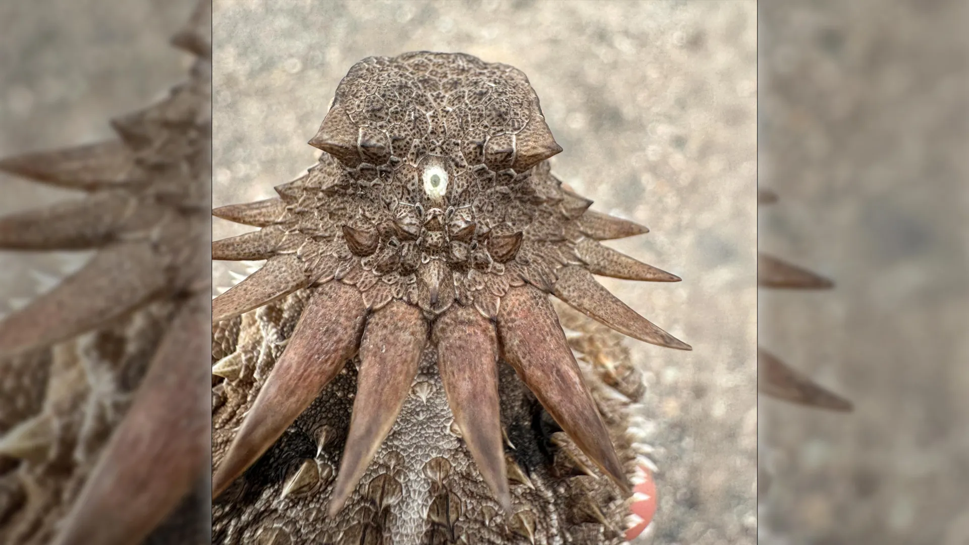
The original median eye retreated inward, sinking deep beneath the skull roof to escape the direct glare of the sun while retaining its profound sensitivity to light. Over hundreds of millions of years, this internalized photoreceptive structure transformed into an endocrine organ: the pineal gland. While humans no longer use the pineal gland to directly "see" light through the skull—as some lower vertebrates, such as certain modern lizards and amphibians, still do with their functional "third eyes"—the gland retains its ancient, ancestral sensitivity to photoperiods. It measures the duration of day and night, translating environmental light signals (relayed via pathways from the modern eyes) into chemical signals, specifically the hormone melatonin.
Supporting Context & Metrics: Decoding the Architectural Divide
To appreciate the gravity of the Lund-Sussex research, one must examine the profound structural gulf separating vertebrate eyes from those of other successful animal phyla.
The Great Evolutionary Divide: Brain vs. Skin
In the grand tapestry of biological design, nature arrived at image-forming eyes along two completely independent developmental pathways:
- The Invertebrate Blueprint (Insects, Mollusks, and Cephalopods): In species such as Drosophila (fruit flies) and Octopus vulgaris, the retina develops from the surface ectoderm—the embryonic tissue that ultimately forms the outer skin of the organism. Cells on the outer surface of the head invaginate or fold inward during embryonic development to form the cup of the eye. Consequently, the photoreceptor cells point outward toward the light source, and the nerve fibers run behind the retina, collecting into the optic nerve.
- The Vertebrate Blueprint (Fish, Amphibians, Reptiles, Birds, and Mammals): In stark contrast, the vertebrate eye emerges directly from the diencephalon region of the embryonic brain. As the neural tube develops, optic vesicles push outward toward the surface of the head. This means the vertebrate retina is quite literally an exposed piece of the central nervous system. A striking anatomical quirk of this developmental origin is that vertebrate retinas are effectively "inside-out": light must pass through layers of transparent nerve cells and blood vessels before finally striking the photoreceptor cells (rods and cones) nestled at the very back of the eye.
| Feature | Vertebrate Eyes (Humans, Fish, Birds) | Invertebrate Eyes (Insects, Squids) |
|---|---|---|
| Embryonic Origin | Central Nervous System (Brain outpocketing) | Surface Ectoderm (Skin/Epidermis) |
| Retinal Architecture | "Inside-out" (Light passes through neural layers first) | Direct (Light strikes photoreceptors immediately) |
| Neural Circuitry | Processing circuits integrated directly within the retina | Processing circuits housed in separate optic lobes of the brain |
| Evolutionary Root | Rebuilt from a median cyclopean ancestor | Evolved directly from surface light-sensitive skin patches |
Unlocking the Neural Circuitry of the Retina
The revelation that vertebrate eyes stem from a centralized median structure also solves a long-standing mystery regarding retinal neurobiology: the origin of the complex neural circuits that pre-process visual data before images ever reach the visual cortex of the brain.
In invertebrate eyes, the neural processing centers (such as the optic lobes) sit as distinct masses behind the eye structure. In vertebrates, however, cells like bipolar cells, horizontal cells, and amacrine cells are tightly packed directly inside the retina. The new research demonstrates that these sophisticated preprocessing networks are direct inheritances from the neural machinery that once operated the ancient median eye. When the median structure expanded to create our modern paired eyes, it brought its sophisticated internal wiring along for the ride, cementing a neuro-developmental architecture unique to our lineage.
Official Statements and Expert Analysis
The implications of this study extend far beyond comparative anatomy, touching upon the fundamental rules governing how complex biological systems evolve over deep time.
Weighing in on the disruptive nature of the findings, Professor Emeritus Dan-E Nilsson of Lund University—one of the world’s leading authorities in sensory biology—emphasized the paradigm-shifting scope of the discovery:
"The results are a surprise. They turn our understanding of the evolution of the eye and the brain upside down," Nilsson stated during the publication of the research.
Reflecting on the strange transitional phases of our deep ancestry, Nilsson noted how the loss and subsequent reconstruction of vision reshaped our anatomical destiny:
"We don’t know whether the paired eyes in our branch of the evolutionary tree were just light-sensitive cells or simple image-forming eyes. We only know that the organism later lost them."
Crucially, Nilsson highlighted how this discovery finally resolves the structural anomaly that has perplexed generations of anatomists studying why vertebrate vision operates so differently from that of the animal kingdom’s other visual heavyweights:
"Now we finally understand why the eyes of vertebrates differ so radically from the eyes of all other animal groups, such as insects and squid. The film of our eyes — the retina — developed from the brain, whereas the eyes of insects and squid originate in the skin on the sides of the head."
Furthermore, Nilsson pointed out the astonishing continuity between ancient environmental adaptations and modern human physiology, bridging the gap between ancient Precambrian worms and modern chronobiology:
"For the first time, we now also understand the origin of the neural circuits that analyze the image in our retina… It’s mind-boggling that our pineal gland’s ability to regulate our sleep according to light stems from the cyclopean median eye of a distant ancestor 600 million years ago."
Collaborating researchers from the University of Sussex echoed these sentiments, noting that advanced molecular phylogenetics and detailed cellular mapping were vital in untangling the web of homologies that connect modern neuro-anatomy to Ediacaran microfossils. By comparing the specific light-transducing proteins (opsins) and structural transcription factors across diverse phyla, the research team successfully mapped out the step-by-step molecular journey that transformed a solitary spot of light-sensitive skin on a worm’s head into the sophisticated neuro-optical machinery that powers human reading, art, and spatial awareness today.
Future Outlook: Implications for Neuroscience and Evolutionary Theory
As the scientific community digests the conclusions put forward by the Lund-Sussex research team, the ripple effects are already being felt across multiple disciplines, promising exciting new avenues for future investigation.
1. Rewriting Evolutionary Textbooks
The traditional view of linear evolutionary progression—where complex organs steadily accumulate refinements over unbroken spans of time—is facing a necessary reckoning. This study provides a striking textbook example of "evolutionary recycling and reconstruction," proving that lineages can completely abandon complex sensory organs, rely on simplified backup systems for millions of years, and then construct entirely novel sensory apparatuses from those minimalist foundations. Evolutionary biologists are now re-examining other complex organ systems—such as the vertebrate inner ear and the central nervous system—to determine whether similar "detours" occurred in their development.
2. Advancing Chronobiology and Sleep Medicine
Understanding the deep evolutionary roots of the pineal gland provides a richer context for human chronobiology. The pineal gland’s regulation of melatonin production in response to environmental photoperiods is intimately tied to human health, influencing everything from seasonal affective disorder (SAD) to circadian rhythm sleep disorders and metabolic regulation. By tracing the cellular ancestry of the pineal gland back to a primitive light-monitoring organ, researchers gain deeper insight into the fundamental signaling pathways that link light exposure directly to endocrine function, potentially guiding novel therapeutic interventions for sleep disturbances in an increasingly artificial, screen-lit modern world.
3. Regenerative Medicine and Neural Engineering
The fact that the vertebrate retina is an direct extension of the brain holds immense promise for regenerative medicine. Neuroscientists working on retinal degenerative diseases, such as retinitis pigmentosa and macular degeneration, frequently encounter the challenge of interfacing artificial implants or stem-cell-derived neural tissues directly with the central nervous system. Understanding the embryonic and evolutionary program that allowed the brain to push outward and form the retina in the first place offers vital clues for bioengineers attempting to repair damaged optic pathways and restore sight to the blind.
Conclusion
The story of vertebrate vision is far stranger and more wondrous than science fiction ever dared imagine. Every time a human being opens their eyes to watch the sunrise, looks up at a starry night sky, or closes their eyes to fall into a deep slumber, they are activating ancient biological machinery forged in the twilight seas of the Precambrian Earth. We are the direct descendants of a tiny, worm-like cyclops that traded its eyes for simplicity, survived in the shadows, and ultimately rebuilt its view of the universe from the inside of its own brain.