
Executive Overview
As winter descends upon the Northern Hemisphere, bringing with it the familiar, unwelcome triad of high fevers, aching limbs, and persistent runny noses, humanity braces for its annual battle against the influenza virus. Despite decades of advanced medical research, seasonal influenza remains a formidable global health challenge, causing millions of cases of severe illness and hundreds of thousands of deaths worldwide every year.
At the microscopic heart of this perennial battle is a deceptively simple biological event: the invasion of a healthy human cell by a viral particle. For generations, virologists have understood the broad strokes of this process—how the pathogen binds to the outside of a cell, maneuvers its way inward, and hijacks the host’s internal machinery to replicate. However, the precise, nanoscopic mechanics of how this violent takeover begins have long remained obscured behind the limitations of traditional scientific technology.
Now, an international collaboration of researchers from Switzerland and Japan has shattered these optical and technological barriers. Under the direction of Yohei Yamauchi, Professor of Molecular Medicine at ETH Zurich, a multidisciplinary research group has unveiled a groundbreaking microscopy approach that allows scientists to zoom in on the outer surface of living human cells in real time and with unprecedented spatial clarity.
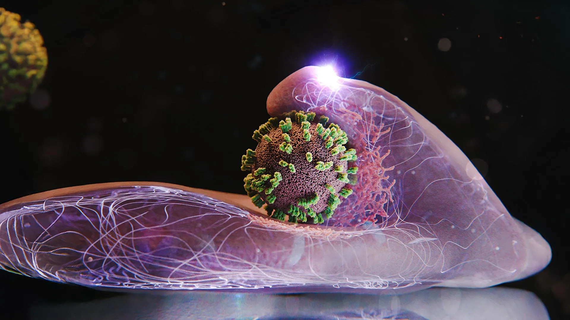
Dubbed ViViD-AFM (virus-view dual confocal and atomic force microscopy), this novel imaging system has enabled researchers to capture the exact moment an influenza virus penetrates a living cell, live and in sharp, high-definition detail. Far from the passive, one-sided invasion scientists once imagined, this dynamic interaction resembles an intricate, synchronized choreography.
The findings upend long-held assumptions about viral entry. Rather than merely serving as helpless victims of an aggressive pathogen, human cells actively participate in the process, unwittingly facilitating their own infection. By exploiting a routine cellular uptake system designed to ingest essential nutrients, the influenza virus effectively "surfs" across the cell membrane, manipulating the host’s structural proteins to gain entry.
This deep dive into the microscopic mechanics of viral infection is more than an academic triumph; it is a paradigm shift for antiviral research. By providing a live, high-resolution window into the earliest stages of viral penetration, ViViD-AFM opens unprecedented avenues for testing novel drug candidates, evaluating vaccine efficacy, and potentially tracking other dangerous pathogens in real time. As researchers look toward the future, this technological leap promises to transform our approach to stopping viral infections before they can even begin.
Detailed Chronology: The Step-by-Step Anatomy of an Influenza Infection
To fully appreciate the breakthrough achieved by Professor Yamauchi and his team, one must trace the chronological sequence of events that occurs when an influenza virus encounters a human cell. Using the newly developed ViViD-AFM technology, scientists have been able to document this microscopic drama frame by frame, revealing a multi-stage process characterized by viral navigation, cellular manipulation, and eventual internal capitulation.
Phase 1: The Arrival and Viral Surfing
The infection cycle begins the moment an influenza virus enters the body, typically via respiratory droplets, and makes contact with the epithelial cells lining the respiratory tract. However, landing on a cell does not immediately guarantee entry. The surface of a human cell is a complex, densely crowded landscape of proteins, lipids, and carbohydrates.
Upon initial contact, the influenza virus does not simply burrow blindly through the membrane. Instead, it engages in what researchers describe as "viral surfing." The virus attaches to specific surface molecules—primarily sialic acid receptors—acting as cellular docking sites.
Using the advanced tracking capabilities of ViViD-AFM, researchers observed that once attached, the virus does not remain stationary. It moves dynamically along the cell’s membrane, latching onto one receptor after another in a fluid, sliding motion. This surface migration is not random; the virus actively navigates toward specialized zones on the cell surface that are densely populated with these receptors. A micro-environment featuring a high concentration of receptors provides the most thermodynamically stable and efficient entry route for the pathogen.
Phase 2: Hijacking the Nutrient Pipeline
One of the most profound discoveries made by the ETH Zurich and Japanese research team is that the cell’s initial response to the surfing virus is remarkably active. Although the host cell gains absolutely no benefit from being infected—and, indeed, faces systemic destruction as a result—the interaction mimics normal, healthy cellular operations.
The influenza virus is an evolutionary master of disguise and exploitation. It survives and propagates by hijacking a routine cellular uptake mechanism known as receptor-mediated endocytosis. This internal pipeline is vital for the cell’s survival, responsible for importing essential life-sustaining substances such as hormones, cholesterol, and iron from the extracellular environment.
Because the cell cannot disable this essential transport system without starving itself, the influenza virus effectively weaponizes it. The virus’s outer surface proteins mimic the chemical signatures of the nutrients the cell actively seeks to ingest, tricking the host into rolling out the welcome mat.
Phase 3: The Clathrin Trap and Membrane Deformation
Once the virus has navigated to a receptor-rich zone and signaled its presence, the mechanical phase of the invasion begins. ViViD-AFM imagery reveals that the cell’s plasma membrane responds by forming a distinct indentation directly beneath the attached pathogen.
This is where a specialized structural protein called clathrin enters the narrative. Upon detecting the viral attachment, the cell summons crucial clathrin molecules to the exact site of the intrusion. These proteins assemble into a polyhedral lattice, shaping and structurally supporting a deepening pocket within the membrane.
As the clathrin-coated pit expands, it actively wraps itself around the influenza virus, enclosing the pathogen within a nascent membrane vesicle. The cell then pulls this vesicle inward, detaching it from the outer membrane and drawing it deep into the cell’s interior. Once safely inside the intracellular environment, the protective protein coat dissolves, releasing the viral genetic material into the cytoplasm, where it can begin hijacking the host’s machinery to replicate.
Phase 4: The Cell’s Active Participation
Perhaps the most astonishing revelation captured by the ViViD-AFM system is that the cell does not merely submit to this process; it actively assists the virus at multiple stages of entry.
When researchers tracked the membrane dynamics in real time, they observed that the cell’s surface pushes upward toward the virus, almost as if trying to reach out and seize it. These wave-like cellular motions intensify dramatically if the virus attempts to drift away from the surface. In essence, the cell’s internal actin cytoskeleton generates forces that inadvertently help pull the virus into the trap, completing a dangerous biological feedback loop where the host works against its own survival.
Supporting Context & Metrics: Overcoming Technological Frontiers
The breakthroughs achieved by Professor Yamauchi’s research group were made possible only by transcending the severe limitations that have historically plagued microscopic virology. For decades, scientists studying viral entry were caught in a persistent technological dilemma: they had to choose between high spatial resolution and real-time observation.
The Limits of Conventional Microscopy
To understand the magnitude of the ViViD-AFM breakthrough, it is necessary to examine the shortcomings of older imaging methodologies:
- Electron Microscopy: For years, electron microscopy served as the gold standard for visualizing nanoscale biological structures. It offers extraordinary spatial resolution, allowing scientists to view individual viral particles and cellular components in minute detail. However, electron microscopy requires rigorous sample preparation—including chemical fixation, dehydration, and heavy-metal coating—which invariably kills the cells. Consequently, electron microscopy can only ever capture isolated, static snapshots in time. It is the microscopic equivalent of trying to understand a feature-length ballet by examining a single photograph of a dancer mid-leap.
- Fluorescence Microscopy: Conversely, traditional fluorescence microscopy allows for live, real-time imaging of cellular processes. Researchers can tag viral particles or cellular proteins with fluorescent markers and watch them move across a living sample. The fatal flaw of this method, however, is its notoriously low spatial resolution. The diffraction limit of light makes it exceptionally difficult to observe the fine-scale, nanometer-level mechanical interactions occurring at the exact membrane interface where a virus enters a cell.
The Birth of ViViD-AFM
To bridge this technological chasm, the research team from Switzerland and Japan engineered a hybrid instrument: virus-view dual confocal and atomic force microscopy (ViViD-AFM).
This innovative approach marries the strengths of two distinct technologies:
- Atomic Force Microscopy (AFM): AFM operates not by using light, but by physically scanning a surface with an ultra-fine, microscopic tip—much like a record player needle reading the grooves of a vinyl disc. This tactile scanning yields topographical maps with nanometer-scale spatial resolution, capturing the physical deformation of cell membranes in real time.
- Confocal Fluorescence Microscopy: Integrated seamlessly with the AFM unit, confocal fluorescence optics provide real-time chemical tracking, allowing researchers to illuminate specific proteins (such as clathrin) with fluorescent tags as they migrate to the site of infection.
By combining atomic-scale physical topography with live fluorescence tracking, ViViD-AFM provides a comprehensive, four-dimensional view of viral entry. Researchers can now observe not just where a virus is, but how the physical membrane deforms, when structural proteins are summoned, and why specific micro-environments dictate the success of an infection.
Official Statements & Expert Perspectives
The implications of this research extend far beyond the confines of the laboratory in Zurich. Experts in molecular medicine and virology are heralding the discovery as a milestone in our understanding of host-pathogen interactions.
"The infection of our body cells is like a dance between virus and cell," explains Yohei Yamauchi, Professor of Molecular Medicine at ETH Zurich and director of the research group.
Elaborating on the psychological and philosophical shift in how virologists view host cells, Yamauchi emphasizes that the traditional model of a passive cell being breached by an aggressive parasite is fundamentally incomplete.
"For years, we pictured viral entry as a purely mechanical breaching of a wall. What our live-imaging setup reveals is an active, highly cooperative dialogue—albeit a fatal one—between the host membrane and the incoming pathogen. The cell is tricked into using its own energy, its own structural proteins, and its own transport mechanisms to pull the enemy inside."
Independent virologists not involved in the study have also praised the technical ingenuity of the ViViD-AFM platform. Dr. Elena Vance, a senior researcher in viral pathogenesis at the European Molecular Biology Laboratory, noted:
"The marriage of atomic force microscopy and confocal fluorescence is a technical tour de force. By allowing us to witness membrane dynamics and protein recruitment simultaneously and in real time, Yamauchi and his team have resolved debates that have stalled our field for over a decade. This is not just a win for influenza research; it is a new lens through which we can study cellular biology as a whole."
Future Outlook: Implications for Antiviral Research and Global Health
As seasonal influenza continues to claim hundreds of thousands of lives globally and poses a perpetual threat of mutating into a pandemic strain, the development of the ViViD-AFM platform arrives at a critical juncture. The applications of this technology extend across multiple pillars of modern medicine, promising to revolutionize how we combat infectious diseases.
1. Direct Screening of Antiviral Drug Candidates
Traditional drug discovery relies heavily on high-throughput biochemical assays that measure whether a compound can inhibit a viral enzyme or block a receptor in a test tube. While effective, these assays often fail to predict whether a drug will succeed in the complex, crowded environment of a living cell.
Because ViViD-AFM allows scientists to observe an infection unfolding in real time within living cell cultures, it provides a powerful, direct testing ground for antiviral drug candidates. Researchers can introduce experimental compounds into the Petri dish and watch instantly whether the drug halts viral surfing, prevents clathrin recruitment, or stops membrane indentation. This real-time visual feedback can dramatically accelerate the identification and optimization of broad-spectrum antivirals.
2. Expanding Horizons: Beyond Influenza
While the initial studies have focused heavily on influenza viruses, the underlying mechanical principles of viral entry are remarkably conserved across many dangerous pathogen families. Viruses such as coronaviruses, Ebola, and HIV all rely on hijacking host cell membrane dynamics to gain entry.
The research team anticipates that the ViViD-AFM technique can be readily adapted to study these diverse pathogens. By mapping the specific entry "dances" of different viruses, scientists may uncover universal vulnerabilities in how pathogens interact with human cells—paving the way for pan-viral therapeutics capable of stopping multiple disease families with a single intervention.
3. Enhancing Vaccine Design and Evaluation
In addition to antiviral therapeutics, the real-time imaging capabilities of ViViD-AFM offer exciting possibilities for vaccine research. Understanding exactly how neutralizing antibodies interfere with viral surfing and membrane penetration at the single-molecule level can help immunologists design more effective, durable vaccines. By visualizing how antibody-coated viral particles are neutralized before they can trigger the clathrin uptake mechanism, researchers can fine-tune vaccine formulations to elicit the most potent protective responses.
Conclusion
The annual return of influenza has long been accepted as an inevitable facet of human life, met each year with seasonal vaccines and supportive care. Yet beneath the macroscopic misery of fevers and fatigue lies a microscopic war zone—a delicate, highly coordinated cellular dance that determines the course of human health.
Through the visionary work of Professor Yohei Yamauchi and his team at ETH Zurich and their Japanese collaborators, humanity now possesses a front-row seat to this microscopic ballet. By pioneering the ViViD-AFM platform, these scientists have shattered old technological boundaries, transforming our understanding of how viruses enter cells from a series of static snapshots into a dynamic, living narrative. As this technology transitions from basic research into applied antiviral development, it carries the profound promise of turning the tables on influenza—giving science the tools not just to treat infection, but to intercept it at the very moment it begins.