
By Brandy Willeford, DVM, MS, DACVS-SA
Executive Overview
Meticulous hemostasis has stood as a foundational principle of veterinary surgery since the inception of modern veterinary education. Achieving precise control over intraoperative bleeding minimizes overall blood loss, curtails unnecessary tissue trauma, preserves clear visualization of the surgical field, and ultimately enhances the efficiency and safety of virtually any procedure. While traditional steel scalpels and manual ligatures remain staples of the operating room, veterinary medicine has increasingly embraced advanced energy-based tools to optimize surgical outcomes.
Electrosurgery stands out as one of the most versatile technologies available to modern veterinary practitioners. Supported by veterinary and human medical studies demonstrating reduced blood loss and shorter operating times, electrosurgical instruments are valuable additions to any progressive surgical suite. However, not all electrosurgical devices operate on the same biological or physical principles. A comprehensive understanding of the operational differences between monopolar, bipolar, and advanced vessel-sealing devices is critical for surgeons seeking to harness these technologies safely and effectively.
This article explores the mechanics of electrosurgery, contrasts it with traditional electrocautery, breaks down the utility and safety considerations of both monopolar and bipolar modalities, evaluates the role of advanced vessel-sealing systems, and offers practical guidance for equipment management in busy veterinary hospitals.
Detailed Chronology and Technical Evolution of Surgical Hemostasis
To appreciate the advent of modern electrosurgery, it is helpful to trace the evolution of hemostatic techniques within surgical practice. Historically, veterinary surgeons relied exclusively on mechanical methods—such as digital pressure, hemostatic forceps, and suture ligatures—to control bleeding. While effective, these techniques can be time-consuming, particularly in diffuse capillary bleeding or when working within deep, confined anatomical spaces.
The introduction of thermal hemostasis revolutionized the surgical field. Initially, this involved electrocautery, a technology frequently confused with electrosurgery. True electrocautery utilizes direct electrical current to heat a resistive metallic instrument (much like a small branding iron), which is then applied to tissue to achieve thermal coagulation. Crucially, in electrocautery, the electrical current itself never passes through the patient’s body.
In contrast, electrosurgery—pioneered and refined over the past century—directs an electrical waveform straight through the patient’s tissues. The high-frequency alternating current interacts directly with cellular fluid and tissue proteins, generating localized heat that either vaporizes cells to cut tissue or denatures proteins to achieve hemostasis.
Over the decades, electrosurgical generators evolved from rudimentary, single-waveform analog units into sophisticated, micro-processor-controlled systems capable of delivering blended waveforms, constant power output across varying tissue impedances, and automated safety monitoring. The late 20th and early 21st centuries saw the introduction of bipolar electrosurgery, which eliminated the need for patient grounding pads by confining current between two closely apposed electrodes. This paved the way for modern vessel-sealing devices, which combine sophisticated bipolar energy delivery with mechanical compression to permanently fuse large vascular structures without traditional suture ligatures.
Supporting Context & Operational Metrics: Monopolar vs. Bipolar Systems
Implementing electrosurgery safely requires a firm grasp of the physics governing the two primary surgical circuits: monopolar and bipolar.
Monopolar Electrosurgery
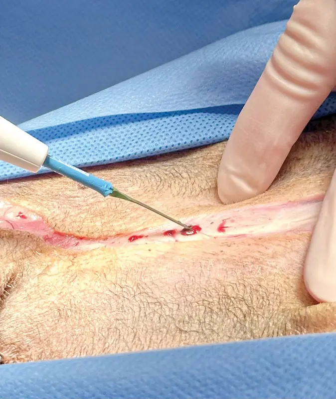
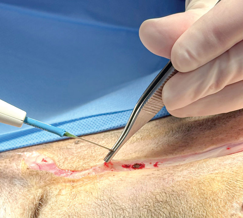
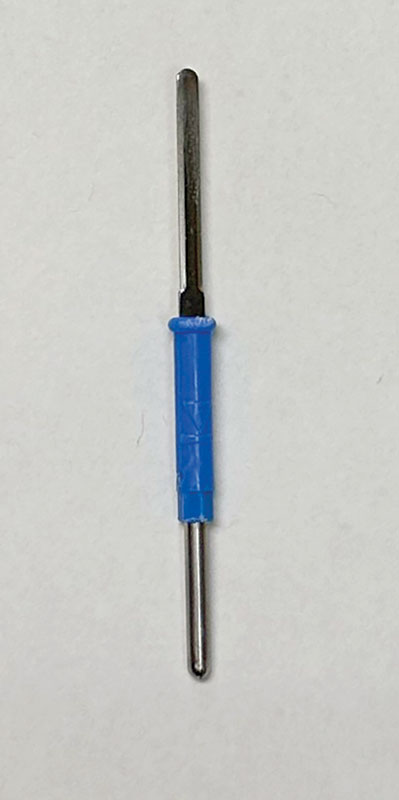
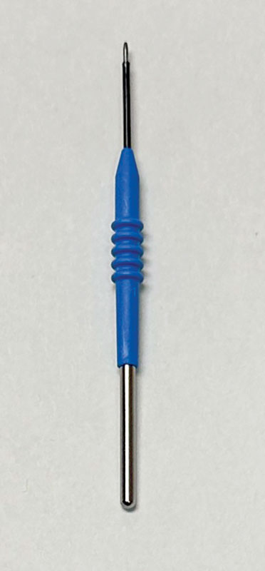
Monopolar electrosurgery is the most widely recognized form of electrosurgery in veterinary clinics. The system relies on a complete circuit: a generator, an active electrode (housed within a pencil-like handpiece), the patient’s body, a dispersive (grounding) pad, and a return path back to the generator.
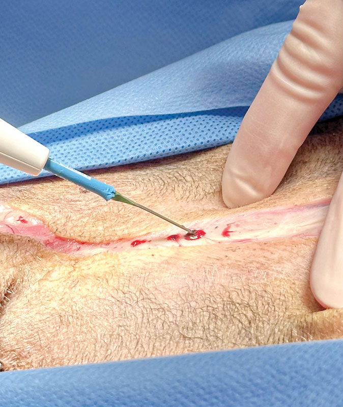
- Cutting Mode: Utilizes a continuous, low-voltage electrical waveform that rapidly heats intracellular water, causing cellular vaporization and clean tissue dissection.
- Coagulation Mode: Utilizes an intermittent, high-voltage waveform that heats tissue in brief bursts, desiccating cells and promoting protein denaturation for effective hemostasis. Coagulation energy can be applied directly to a bleeding point or directed through a metal instrument (coaptive coagulation) to occlude larger vessels.
Operational Metrics and Safety Considerations
Despite its efficiency, monopolar electrosurgery demands rigorous attention to patient preparation and environmental safety:
- Fluid Interference: Monopolar energy is less effective in a wet surgical field because pooling fluids can divert the electrical current away from the target tissue.
- Dispersive Pad Placement: Correct application of the grounding pad is non-negotiable. Pads must maintain complete contact with the patient’s skin, utilizing conductive gel when specified by the manufacturer. Poor pad contact concentrates electrical return current, risking thermal skin burns.
- Insulation and Patient Positioning: Patients must be strictly insulated from metal surgical tables. All conductive materials—such as metal collars, harnesses, or monitoring leads—must be removed to prevent unintended alternate current pathways.
- Fire Safety Hazards: Alcohol-based skin preparation solutions are highly flammable. Electrosurgical activation in the presence of pooled alcohol or residual vapors can spark operating room fires. Prep solutions must be allowed to dry completely before draping and activating any energy device.
+-----------------------------------------------------------------+
|                  MONOPOLAR ELECTROSURGERY CIRCUIT               |
|                                                                 |
|  [Generator] ---> [Active Electrode] ---> [Patient Tissue]      |
|         ^                                           |           |
|         |                                           v           |
|  [Return Path] <--- [Dispersive Pad] <--------------+           |
+-----------------------------------------------------------------+
Bipolar Electrosurgery
Bipolar electrosurgery addresses several limitations of monopolar systems by incorporating both the active and return electrodes into a single instrument, typically resembling a pair of forceps.
Electrical current travels strictly through the localized tissue grasped between the two tips. Because the current path is short and self-contained, a dispersive grounding pad is entirely unnecessary. This configuration makes bipolar electrosurgery exceptionally useful when working in wet surgical fields or performing delicate dissections near critical structures, such as nerves or major vessels. The primary trade-off is that coagulation times may be slightly longer compared to monopolar devices, depending on tissue impedance and generator settings.
Advanced Vessel-Sealing Technology: Evidence and Application
Vessel-sealing devices represent an advanced subclass of bipolar electrosurgery. By applying a computer-regulated combination of mechanical compression and precise bipolar electrical energy, these devices denature collagen and elastin within vessel walls, permanently fusing the vessel lumen.
Clinical Efficacy and Anatomical Limitations
While many commercial vessel-sealing units are marketed for sealing vessels up to 7 mm in diameter, surgeons must never assume uniform performance across all tissue types. Burst-pressure studies and clinical evaluations provide vital boundaries for safe usage:
- Uterine Horns and Bodies: Research evaluating vessel-sealing devices against traditional encircling ligatures in canine uterine horns and bodies demonstrated comparable burst pressures in structures under 9 mm in diameter. However, seals performed on structures 9 mm or larger exhibited significantly lower burst pressures. Consequently, evidence-based guidelines advise against relying solely on vessel sealers for uterine structures exceeding 9 mm.
- Cystic Ducts: In a retrospective study of 47 dogs undergoing open cholecystectomy for gallbladder mucoceles, researchers evaluated the perioperative and short-term complications of securing the cystic duct with a vessel-sealing device. The findings confirmed that the technique is safe and effective within that specific patient population.
The Economics and Safety of Device Reuse
Many vessel-sealing handpieces are labeled by manufacturers as single-use devices. However, economic pressures in veterinary practice often lead hospitals to re-sterilize and reuse these instruments. This practice introduces mechanical and electrical risks.
Scientific evaluations tracking the repeated sterilization of popular vessel-sealing devices revealed distinct failure thresholds:
- Small Scissor-Style Handpieces: Demonstrated structural or functional failure after a mean of 17.2 sterilization cycles (with the earliest failure observed at 10 cycles).
- Large Handpieces: Failed after a mean of 20 cycles (with the earliest failure occurring at 17 cycles).
To mitigate intraoperative failure, clinics that choose to reuse these instruments should institute strict tracking protocols. Authors recommend discarding small scissor-style handpieces after no more than nine sterilization cycles, and larger handpieces after 16 cycles—or immediately discarding any device showing early signs of mechanical wear or performance degradation.
Future Outlook: Technology, Training, and Best Practices
As veterinary surgery continues to advance, electrosurgical technology will undoubtedly become even more sophisticated, integrating tissue-sensing feedback loops that automatically adjust energy delivery based on real-time changes in tissue impedance. However, technology should never replace surgical judgment.
Future integration of electrosurgery in veterinary hospitals relies on three core pillars:
- Comprehensive Training: Veterinary teams must understand the fundamental biophysics governing energy-tissue interactions, recognizing why monopolar systems demand meticulous grounding while bipolar and vessel-sealing systems operate locally.
- Standardized Safety Protocols: Clinics must enforce rigorous protocols regarding alcohol dry times, grounding pad placement, and table insulation to eliminate preventable patient injuries.
- Evidence-Based Equipment Utilization: Surgeons must respect the validated physical limits of energy devices—such as maximum vessel diameters and sterilization cycle limits for single-use instruments—to ensure optimal patient safety.
Ultimately, electrosurgery is simply another instrument in the veterinary surgeon’s armamentarium. Like a scalpel, needle holder, or suture needle, its true clinical value depends entirely on the surgeon’s working knowledge of when to apply it, how to operate it safely, and when traditional techniques remain the superior choice.
References
- Studies comparing blood loss in veterinary and human medicine tissue incisions (referenced in text).
- Comparative analyses of scalpel vs. electrosurgical cutting efficiency (referenced in text).
- Surgical time reduction metrics utilizing advanced electrosurgical modalities (referenced in text).
- Biophysical principles of tissue vaporization, protein denaturation, and collagen/elastin fusion (referenced in text).
- Burst pressure evaluations of vessel-sealing devices versus encircling ligatures in canine reproductive tracts.
- Retrospective evaluations of vessel-sealing device safety during canine cholecystectomy for gallbladder mucocele.
- Material degradation and mechanical failure studies of re-sterilized single-use vessel-sealing handpieces.