
Executive Overview
Feline eyes have long captured the human imagination, inspiring centuries of folklore, poetry, and artistic fascination. With their remarkable ability to dilate in the dark and narrow into precise, vertical slits under bright illumination, a cat’s eyes are both masterworks of evolutionary design and windows into their mysterious inner lives. Yet, even among seasoned veterinary professionals who spend decades examining the intricate anatomy of domestic animals, a rare anatomical anomaly can still stop an experienced clinician in their tracks.
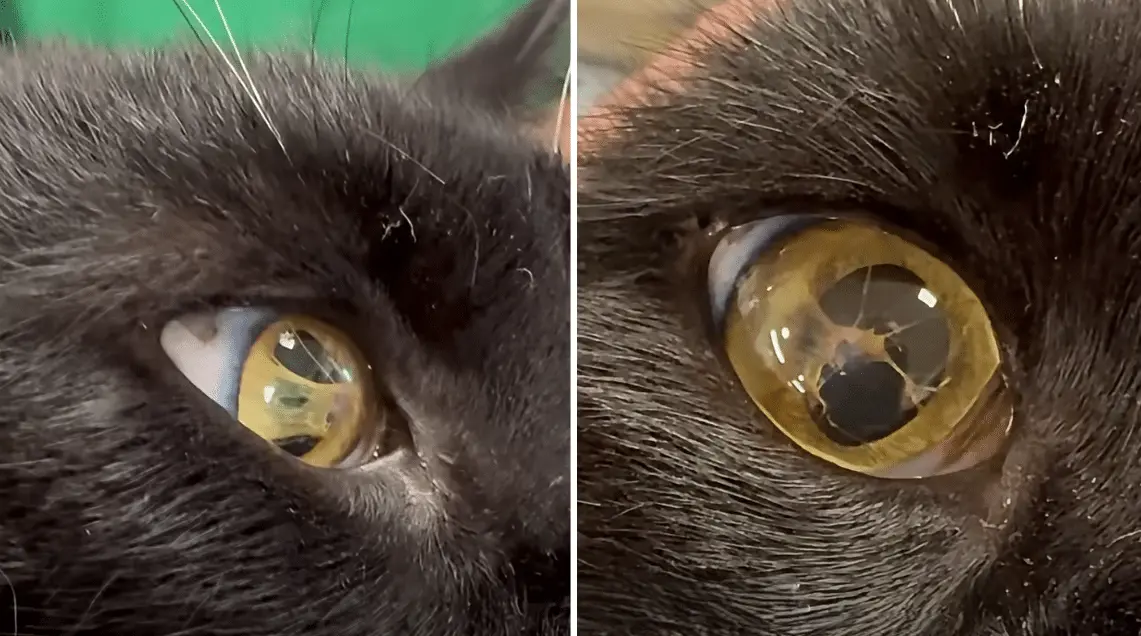
This exact scenario unfolded recently at the Jarrettsville Vet Center, where a routine examination of a striking black cat revealed an extraordinary feature hidden within the animal’s eye. Rather than immediately publishing a clinical diagnosis, the veterinary team decided to leverage social media, transforming the unusual discovery into an interactive educational puzzle for their online community. By inviting followers to closely inspect the cat’s eye and guess the nature of the anomaly, the clinic sparked a widespread conversation about a little-known congenital condition.
The answer to the veterinary riddle was persistent pupillary membrane (PPM), a fascinating and rarely discussed developmental occurrence rooted in the earliest stages of feline embryology. While the clinical terminology sounds formidable, the reality is a nuanced natural variation that offers a remarkable glimpse into the complex biology of prenatal growth. This in-depth investigative report examines the mechanics of persistent pupillary membrane in cats, exploring its embryological origins, potential impacts on feline vision, necessary diagnostic protocols, and the broader implications of these rare veterinary encounters in modern clinical practice.
Detailed Chronology: From Social Media Mystery to Veterinary Diagnosis
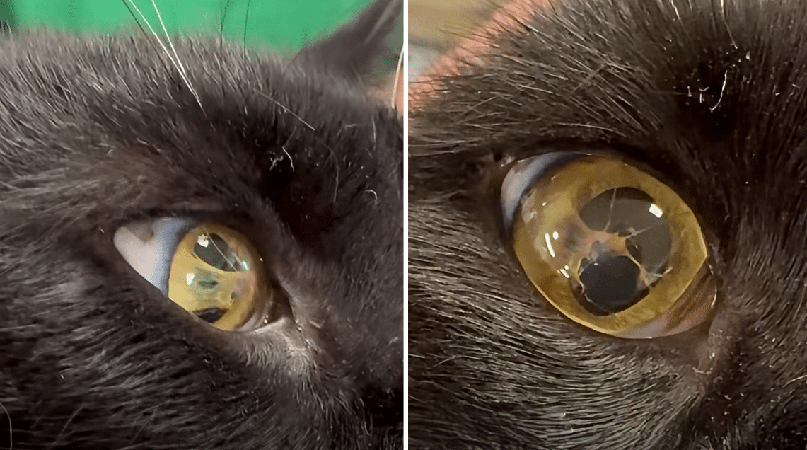
The unfolding of this veterinary curiosity began inside the examination rooms of the Jarrettsville Vet Center, an established animal care facility known for blending compassionate patient treatment with engaging public outreach. During a standard wellness visit, a sleek black cat presented with an unmistakable visual feature inside one of its eyes—a subtle, web-like structure spanning the pupil that immediately set it apart from the average feline patient.
Recognizing the unique educational potential of the case, the veterinary staff documented the feature on video. Instead of broadcasting the medical term right away, the clinic uploaded the footage to its official Facebook page, issuing a playful challenge to its followers: take a close look, examine the anomaly, and guess what was happening inside the feline’s eye. The post quickly captured the attention of animal lovers, veterinary students, and fellow clinicians alike, generating a flurry of speculative comments and hypotheses.
After allowing the online community time to analyze the footage, the Jarrettsville Vet Center staff revealed the definitive diagnosis in the comment section: a persistent pupillary membrane. This strategic approach achieved two distinct goals. First, it highlighted a rare congenital eye condition in an engaging, approachable manner. Second, it demonstrated how everyday veterinary medicine frequently intersects with rare biological phenomena, turning an ordinary check-up into an interactive masterclass in feline anatomy.
Supporting Context & Metrics: Understanding Feline Ocular Embryology
To fully comprehend what a persistent pupillary membrane is, one must first look backward in time—specifically, to the developmental stages a kitten undergoes inside the womb long before its eyes ever open to the world.
The Embryonic Life Cycle of the Feline Eye
During prenatal development, a kitten’s rapidly growing eyes require a dedicated source of nourishment and oxygen to support the formation of complex intraocular structures. To achieve this, nature deploys a temporary, specialized network of blood vessels and connective tissue known as the pupillary membrane. This delicate biological scaffolding stretches across the aperture of the pupil, nourishing the developing lens during the gestational period.
As a kitten approaches birth and prepares to transition into a world of light and shadow, this temporary network has a specific biological mission to complete: it must regress, dissolve, and disappear entirely. By the time a healthy kitten opens its eyes weeks after birth, the pupillary membrane has normally vanished, leaving the pupil completely clear and unobstructed to regulate light entry.
When Development Leaves Remnants: The Mechanics of PPM
In rare instances, however, biological regression is incomplete. Tiny remnants of the embryonic pupillary membrane fail to dissolve, remaining anchored inside the fully formed eye. These lingering threads, strands, or web-like membranes constitute what veterinarians define as a persistent pupillary membrane (PPM).
The structural manifestations of PPM vary widely from one animal to the next:
- Iris-to-Iris Strands: The most common variation, where delicate threads stretch from one part of the colored iris across the pupil to another section of the iris.
- Iris-to-Lens Attachments: Fibrous strands extending from the iris and attaching directly to the crystalline lens of the eye.
- Iris-to-Cornea Adhesions: Less common strands that bridge the gap between the iris and the inner endothelial surface of the cornea (the clear outer window of the eye).
While PPM can occur in various domestic species—including dogs, horses, and cats—it is considered uncommon in felines, which explains why the Jarrettsville Vet Center patient immediately captivated veterinary professionals and cat enthusiasts alike.
Clinical Implications: Vision, Diagnosis, and Treatment
When pet owners encounter a diagnosis with a complex medical name like persistent pupillary membrane, their immediate concern invariably centers on their companion’s comfort and visual capability. Fortunately, clinical data and veterinary ophthalmology guidelines provide reassuring context.
Impact on Feline Vision
Whether a cat with PPM experiences vision impairment depends entirely on three key factors: location, density, and extent of the remaining tissue.
- Mild and Benign Cases: In the vast majority of feline cases, PPM strands are exceptionally fine, wispy, and transparent or pigmented. If these strands stretch harmlessly across the iris without interfering with the visual axis, the cat’s vision remains entirely normal. The animal experiences no pain, irritation, or functional deficit.
- Complex and Complicated Cases: When persistent membranes are heavier, or when they anchor aggressively to the crystalline lens or the cornea, secondary complications can arise. Attachments to the lens can occasionally result in localized opacities (small cataracts) or pigment spots on the lens capsule. Similarly, strands adhering to the cornea can cause localized corneal edema (cloudiness). In these more pronounced scenarios, minor visual deficits or light scattering may occur.
Diagnostic Protocols: The Role of the Veterinary Ophthalmologist
Because an unusual eye appearance can stem from multiple distinct etiologies—ranging from harmless congenital anomalies to progressive inflammatory diseases like uveitis—professional diagnostic evaluation is indispensable.
Veterinarians utilize specialized equipment, such as direct and indirect ophthalmoscopes and slit-lamp biomicroscopes, to magnify and illuminate the intraocular structures. A slit-lamp examination allows the clinician to view the eye in optical sections, precisely mapping the origin, path, and termination point of any persistent strands. This sophisticated imaging helps differentiate true congenital PPM from post-inflammatory synechiae (adhesions caused by previous eye injuries or feline inflammatory diseases).
Management and Treatment Strategies
For the vast majority of cats diagnosed with mild PPM, no medical or surgical treatment is required. Because the condition is congenital and non-progressive—meaning the leftover strands will not multiply, spread, or behave like an infectious disease—veterinarians typically recommend nothing more than routine monitoring during standard annual wellness exams.
Surgical intervention is exceedingly rare in veterinary ophthalmology and is reserved strictly for severe cases where dense membranes significantly obstruct vision, cause progressive corneal cloudiness, or induce secondary cataracts that severely compromise the animal’s quality of life. For the black cat featured in the Jarrettsville Vet Center video, the physical anomaly stood as a fascinating anatomical signature rather than a medical emergency, reminding the public that nature often leaves unique, harmless artistic touches on her creations.
Future Outlook: Awareness, Education, and Veterinary Medicine
The viral success of the Jarrettsville Vet Center’s social media post highlights a broader, highly positive trend in modern veterinary medicine: the fusion of clinical practice with public digital education. By turning a complex anatomical curiosity into an accessible learning moment, veterinary clinics are bridging the communication gap between specialized medical science and everyday pet guardianship.
Empowering Cat Owners Through Knowledge
For pet owners, understanding conditions like persistent pupillary membrane fosters a deeper appreciation for feline biology. It teaches the vital distinction between harmless anatomical variations and critical red-flag symptoms. While a congenital strand of tissue is benign, cat owners must remain vigilant for true ocular emergencies, such as:
- Sudden squinting or blepharospasm
- Ocular discharge (clear, yellow, or green)
- Persistent redness of the conjunctiva
*明显的 (noticeable) corneal cloudiness or a sudden change in eye color - Behavioral signs of vision loss, such as bumping into furniture or hesitancy in dim lighting
By cultivating observational skills through educational campaigns, veterinary centers empower owners to seek professional care at the earliest sign of true pathology, ensuring better long-term health outcomes for feline companions worldwide.
The Ever-Surprising World of Veterinary Science
Ultimately, the story of this remarkable black cat underscores the endless fascination inherent in veterinary medicine. Even in an era of advanced diagnostics, genetic mapping, and high-resolution imaging, animals continue to surprise clinicians with the sheer diversity of their biological forms. Whether it is an extra toe, a unique ear set, or a delicate web of embryonic tissue suspended across a golden or green iris, these variations remind us of the intricate evolutionary tapestry shared by humans and animals.
As veterinary practices continue to embrace digital platforms for community engagement, educational mysteries like the Jarrettsville Vet Center’s PPM revelation will continue to enrich the public’s understanding of animal health. They remind us that beneath the surface of every ordinary wellness check lies a world of microscopic wonder—proving once again that feline eyes are not only among the most beautiful features in the animal kingdom, but also among the most endlessly captivating.