
Executive Overview
Discovering a cutaneous or subcutaneous mass on a patient is one of the most common, yet clinically complex, scenarios encountered in small animal practice. Whether identified incidentally during a routine annual wellness exam or brought to light by an anxious pet owner who noticed a sudden change at home, the immediate aftermath triggers a cascade of anxious inquiries: What is it? Should we be worried? What do we do next?
For veterinary professionals, managing lumps and bumps is a daily routine. For pet owners, however, these moments are often fraught with intense emotional weight. Pets are cherished family members, and the uncertainty surrounding a new growth can cause profound distress. Recent data underscores this emotional vulnerability, reporting that 21 percent of pet owners consider the death of their pet to be the most distressing loss they have ever experienced. This deep human-animal bond explains why uncertainty regarding a mass feels so burdensome, and why early, practical, and economically accessible diagnostics are paramount.
Dr. Sue Ettinger, DVM, DACVIM (Oncology)—a renowned veterinary oncologist at Guardian Veterinary Specialists in New York and pioneer of the “See Something, Do Something, Why Wait? Aspirate®” initiative—emphasizes a foundational truth in veterinary medicine: no clinician, general practitioner, or veterinary oncologist can look at or palpate a mass and definitively identify its histopathologic nature.
To bridge the gap between uncertainty and actionable diagnosis, the veterinary field is experiencing a technological evolution. Artificial intelligence (AI)-supported digital cytology is rapidly transforming how clinics approach early-stage diagnostics. By delivering real-time, point-of-care cytological insights within approximately 20 minutes, this emerging technology empowers veterinarians to transition clinical conversations from passive monitoring to active investigation, ensuring faster interventions and improved clinical outcomes.
Detailed Chronology: From Clinical Presentation to Surgical Resolution
To fully appreciate the real-world impact of AI-supported digital cytology in modern veterinary practice, it is valuable to examine a clinical case managed by Dr. Ettinger.
Case Study: Zoe’s Diagnosis and Treatment Journey
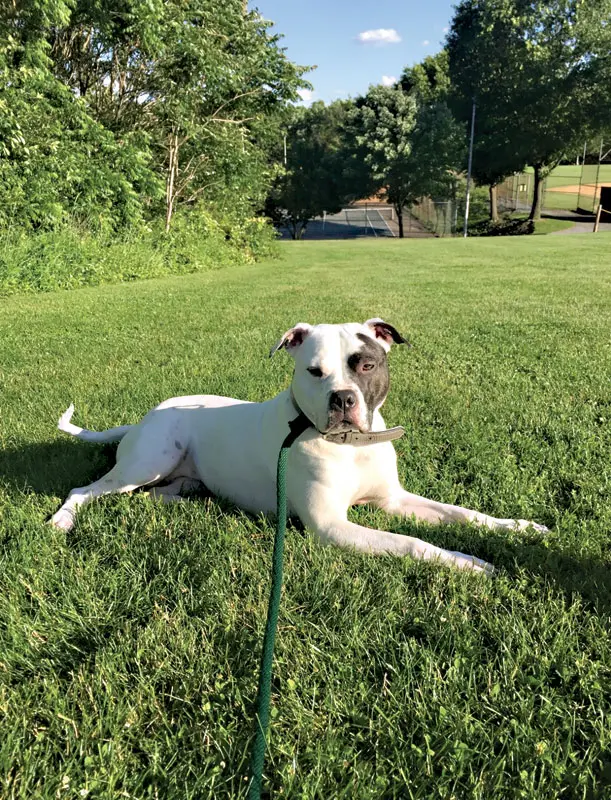
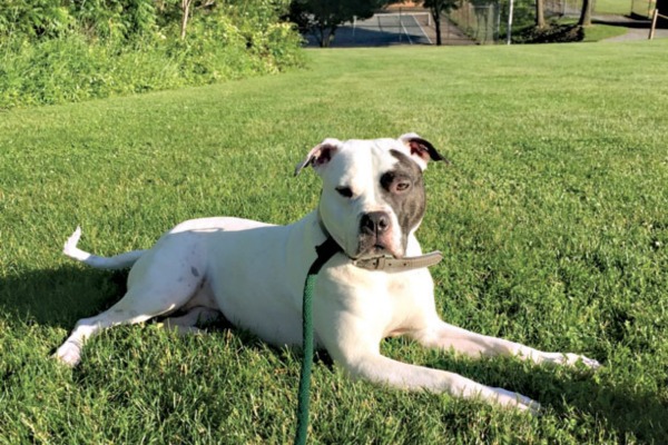
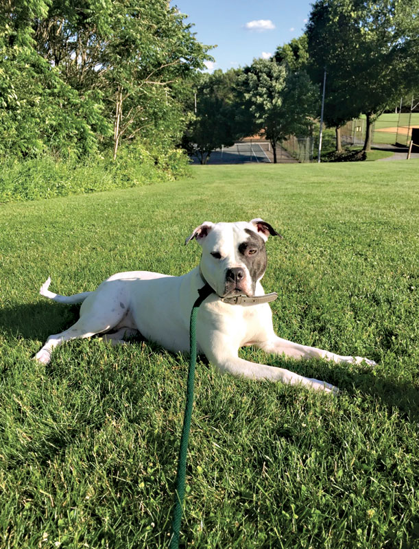
Zoe, an eight-year-old spayed female American Bulldog, presented to Dr. Ettinger for a routine recheck following a previously diagnosed follicular lymphoma with salivary gland involvement. This case represented an atypical presentation of lymphoma; unlike more traditional forms of the disease, Zoe had not undergone a conventional multi-agent chemotherapy protocol following the initial lymph node removal.
1. Initial Presentation and Physical Examination
During the physical examination, Zoe’s peripheral lymph nodes appeared completely normal. However, a clinician’s thorough hands-on assessment revealed a new, flat, movable subcutaneous mass located on the left ventrolateral abdomen.
2. Fine-Needle Aspiration and AI-Supported Screening
Given the discovery of the new mass, Dr. Ettinger performed a fine-needle aspiration (FNA). Clinically, the mass closely resembled a benign lipoma. Furthermore, the lesion’s flat and highly mobile nature made sample acquisition technically challenging.
Rather than sending the slide to an external reference laboratory and forcing the owner to wait days for results, the sample was processed using point-of-care AI-supported digital cytology. Within minutes, the digital platform flagged cell populations inconsistent with a simple lipoma, instead identifying findings highly suggestive of a mast cell tumor (MCT)—the most common malignant cutaneous cancer in dogs.
3. In-Person Consultation and Expert Review
Having actionable cytology results available while the owner was still in the exam room fundamentally changed the trajectory of the visit. Instead of sending Zoe’s family home with instructions to wait anxiously for days, Dr. Ettinger was able to immediately discuss the implications of a mast cell tumor in person.
To ensure absolute diagnostic precision, Dr. Ettinger utilized an add-on expert pathologist review of the digital cytologic grading. This secondary evaluation confirmed a low-grade mast cell tumor, guiding subsequent therapeutic decision-making.
4. Surgical Intervention and Histopathology
Because the mass was subcutaneous and situated on the trunk, Dr. Ettinger recommended surgical excision rather than an intratumoral injection. Eight days post-aspirate, Zoe underwent surgical removal of the mass performed by her primary care veterinarian.
Definitive histopathology confirmed a low-grade subcutaneous mast cell tumor characterized by:
- No infiltrative growth pattern
- Absence of multinucleated cells
- A mitotic count of 0
- Narrow lateral margins, but complete deep margins with a healthy tissue plane beneath the tumor
- Concomitant removal of a separate ventral thoracic lipoma
5. Post-Operative Recheck and Prognosis
Zoe returned to Dr. Ettinger for a post-operative oncology recheck to review the biopsy report in detail. The consultation highlighted that subcutaneous mast cell tumors possess distinct biological behavior compared to dermal mast cell tumors, frequently carrying a highly favorable prognosis when completely excised—though a small subset can exhibit more aggressive tendencies. Additional prognostic assays were discussed but ultimately declined by the owner, with the provision that they could be pursued later if clinically indicated.
Zoe’s case illustrates the seamless integration of AI-supported screening into a complex oncology patient’s timeline, eliminating diagnostic lag and accelerating the path from initial detection to curative surgery.
Supporting Context & Metrics: The Paradigm Shift in Diagnostics
The Philosophy of Early Detection
Dr. Ettinger’s widely adopted “See Something, Do Something, Why Wait? Aspirate®” program was designed to dismantle the historical culture of complacency surrounding cutaneous and subcutaneous masses. The guidelines are purposefully straightforward: if a mass reaches the size of a pea (approximately 1 cm) and has persisted for one month, perform a fine-needle aspiration.
The objective of this initiative is not to induce panic over every minor skin bump, but rather to eradicate the dangerous habit of "wait-and-see" monitoring. Cytology provides a direct window into cellular architecture, differentiating benign inflammatory processes and benign neoplasms from aggressive malignancies before local invasion or metastasis occurs.
Overcoming Traditional Diagnostic Barriers
Historically, cytology faced several operational roadblocks in general veterinary practice:
- Turnaround Times: External reference laboratories typically require 24 to 72 hours (or longer over weekends) to return cytological interpretations, leaving pet owners in an agonizing limbo of uncertainty.
- Financial Constraints: The added costs of courier fees, external pathologist surcharges, and multi-test panels can deter budget-conscious pet owners from pursuing diagnostics.
- Access to Specialists: General practices located in rural or underserved geographic regions may lack rapid access to board-certified clinical pathologists.
The Rise of Artificial Intelligence in Veterinary Medicine
The integration of technology is rapidly altering this landscape. Industry metrics indicate that nearly 40 percent of veterinary professionals in the United States currently incorporate artificial intelligence into their everyday clinical workflows.
AI-supported digital cytology serves as an efficient screening tool at the point of care. Once a stained glass slide is digitally scanned via specialized clinic hardware, proprietary algorithms process the cellular imagery and generate a preliminary report in approximately 20 minutes.
This rapid turnaround allows the veterinary team to evaluate cell populations, differentiate inflammation from neoplasia, and initiate meaningful client conversations during the original appointment. Furthermore, studies show that early diagnosis of malignant tumors—such as mast cell tumors—while they are still small significantly improves long-term prognosis and curability.
Official Statements and Expert Perspectives
The integration of artificial intelligence into oncology diagnostics has sparked vital industry discussions regarding the intersection of advanced technology and human clinical judgment.
The Essential Role of Veterinary Expertise
Veterinary leaders and pathologists universally emphasize that AI-supported tools are designed to augment, not replace, the clinical acumen of licensed veterinarians. Cytology remains an inherently interpretive discipline heavily reliant on clinical context.
"AI-supported digital cytology does not replace veterinarian judgment," explains Dr. Ettinger. "The report still needs to be interpreted in the context of the patient, physical exam, sample quality, and what was seen on the slide. Some cases are straightforward enough to guide the next step, while others are more complex or ambiguous. In those other cases, a clinical pathologist’s expert review can be especially valuable."
Bridging the Gap in General Practice
For general practitioners—who stand on the front lines evaluating dozens of skin masses every week—AI tools provide an essential bridge between initial physical examinations and specialist-level pathology. By streamlining diagnostic workflows, clinics can elevate their standard of care, reduce diagnostic delays, and foster transparent, highly empathetic client communication.
Impact on Client-Veterinarian Trust
Eliminating the agonizing multi-day wait for initial test results fundamentally alters the psychological experience of pet ownership. When a veterinarian can look an owner in the eye and say, "We ran the slides right here in the clinic; our initial screening points toward a mast cell tumor, here is what that means, and here is our immediate plan," the dynamic shifts from helpless anxiety to empowered partnership.
Future Outlook: The Next Frontier in Veterinary Oncology
As artificial intelligence continues to mature, its integration into veterinary medicine will expand beyond basic cytological screening. Future advancements are projected to refine predictive analytics, enhance digital grading algorithms, and streamline tele-cytology networks that connect general practitioners with board-certified oncologists and pathologists in real-time.
However, the core tenets of veterinary medicine will remain unchanged. Early cancer detection will never rely on a single standalone test or piece of software. It will forever depend on a comprehensive triad:
- Careful physical examination and palpation by attentive veterinary professionals.
- Appropriate utilization of diagnostics, reinforced by tools like AI-supported digital cytology.
- Compassionate, transparent communication paired with sound clinical judgment.
By combining cutting-edge diagnostic technology with unwavering clinical expertise, the veterinary community can successfully transition routine lump-and-bump exams from moments of fearful hesitation into opportunities for decisive, life-saving action.
About the Author
Sue Ettinger, DVM, DACVIM (Oncology), is a practicing veterinary oncologist at Guardian Veterinary Specialists in New York. Recognized as the 2019 Western Veterinary Conference Small Animal Continuing Educator of the Year, Dr. Ettinger is a nationally recognized thought leader who pioneered the “See Something, Do Something, Why Wait? Aspirate®” campaign to champion early cancer detection in companion animals. Dedicated to public education, she actively engages with a community of over 140,000 followers across social media platforms and serves as the co-author of the Second Edition of The Dog Cancer Survival Guide.