
By Kendra Freeman, DVM, MS, DACVS (Large Animal/Small Animal)
Specialized Clinical Report & Continuing Education Case Study
Executive Overview
Pyometra—a severe, life-threatening uterine infection characterized by systemic inflammation and purulent fluid accumulation—remains one of the most critical surgical emergencies encountered in veterinary practice. Despite decades of advanced canine reproductive management, educational outreach, and accessible spay (ovariohysterectomy) procedures, intact female dogs continue to succumb to this insidious pathology.
This case study examines a classic, high-stakes presentation: a nine-year-old intact female Bulldog presenting with hallmark signs of lethargy, systemic toxicity, and abdominal discomfort. Through a rigorous examination of the patient’s clinical presentation, clinicopathologic aberrations, and diagnostic imaging findings, this report explores the underlying physiological mechanisms driving the condition.
Furthermore, this article addresses fundamental clinical questions regarding pyometra epidemiology, risk factors such as hormonal exposure and advanced age, and primary bacteriological agents. By blending emergency veterinary protocols with advanced clinical education, this overview aims to arm practitioners with the diagnostic precision and surgical urgency required to manage this systemic crisis successfully.
Detailed Chronology: Case Presentation and Diagnostic Workup
Initial Triage and Clinical Presentation
A nine-year-old intact female Bulldog was presented to the emergency service with a primary complaint of progressive lethargy, acute reluctance to move, and a significant, multi-day decrease in appetite. According to the owner, the dog had completed an estrus cycle approximately six weeks prior, placing her squarely within the high-risk metestrus window for uterine complications.
Upon initial physical examination, the patient was quiet, alert, but dull in demeanor. Vital parameters revealed mild tachycardia and a low-grade fever ($103.1^circtextF$ / $39.5^circtextC$). Mucous membranes were visibly pale and tacky to the touch, indicating early-stage dehydration and compromised peripheral perfusion.
Palpation of the abdomen elicited an immediate, painful response, characterized by marked visceral guarding and a severely tense abdominal wall. Given the signalment—an aging, nulliparous intact female—pyometra immediately shifted to the top of the differential diagnosis list, alongside acute pancreatitis, gastrointestinal perforation, and uroabdomen.
Clinicopathologic Findings
Venous blood was drawn immediately for a complete blood count (CBC), serum biochemistry profile, and electrolyte panel. The results confirmed a profound systemic inflammatory response and secondary metabolic compromise:
- Complete Blood Count (CBC): Demonstrated a marked leukocytosis ($32,500/mutextL$; reference range: $6,000–17,000/mutextL$) characterized by a severe neutrophilia ($28,000/mutextL$) with a regenerative left shift, including toxic bands and metamyelocytes. This picture underscores an overwhelming systemic demand for neutrophils to combat intrauterine infection.
- Serum Biochemistry: Revealed significant azotemia, with elevated blood urea nitrogen (BUN: $52text mg/dL$) and creatinine ($2.4text mg/dL$), indicative of prerenal azotemia secondary to dehydration, compounded by early nephrogenic injury driven by bacterial endotoxemia.
- Protein Panel: Highlighted a striking hyperglobulinemia ($4.8text g/dL$), reflecting chronic antigenic stimulation and systemic antibody production secondary to the persistent intrauterine infection.
Diagnostic Imaging
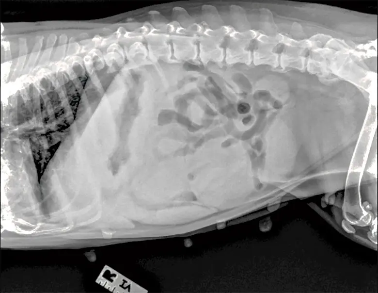
Abdominal radiography was performed to evaluate the architecture of the abdominal cavity and screen for abnormal fluid-filled tubular structures within the visceral space.
As illustrated in the lateral abdominal radiographic view (Figure 1), lateral projections revealed prominent, tortuous, fluid-dense tubular structures throughout the central and caudal abdomen, displacing the intestinal loops craniodorsally. These radiographic anomalies represented the massively distended, fluid-filled uterine horns characteristic of advanced cystic endometrial hyperplasia (CEH)-pyometra complex.
[Lateral Abdominal Radiograph Assessment]
==================================================
Patient: 9-Year-Old Intact Female Bulldog
Findings:
 - Fluid-dense tubular structures visible caudally
 - Craniodorsal displacement of intestinal loops
 - Absence of free peritoneal gas (ruling out rupture)
 - Uterine horn diameter > normal limits (> 1.5 cm)
==================================================
Armed with clinical, hematological, and radiographic confirmation, the surgical team stabilized the patient with aggressive intravenous fluid therapy (balanced electrolyte solutions), initiated broad-spectrum intravenous antimicrobial therapy, and prepared the patient for emergency exploratory laparotomy and ovariohysterectomy.
Supporting Context & Metrics: Epidemiology, Pathophysiology, and Bacteriology
To thoroughly understand the gravity of cases like this Bulldog, veterinary professionals must explore the broader epidemiological and microbiological metrics governing canine pyometra.
1) Risk Factors for the Development of Pyometra
Pyometra is rarely an isolated spontaneous event; rather, it is the terminal phase of a complex endocrinological and immunological cascade.
- Advanced Age: The risk of developing pyometra increases exponentially with age. Repeated exposure to progesterone over successive estrus cycles induces progressive endometrial proliferation and cystic changes (cystic endometrial hyperplasia or CEH). Consequently, older, nulliparous females are at the highest statistical risk.
- Hormonal Administration: Exogenous estrogen administration—historically used for mismatched breeding termination or dermatological conditions—sensitizes the myometrium and upregulates progesterone receptors, exacerbating the risk of secretory phase infections. Furthermore, progestagen compounds utilized for estrus suppression are potent instigators of CEH and subsequent pyometra.
- Breed Predisposition: While any intact female can develop pyometra, epidemiological studies highlight increased relative risk in specific breeds, including Golden Retrievers, Miniature Schnauzers, Rottweilers, Bernese Mountain Dogs, and English Bulldogs.
Correct Answer to Question 1: d) All of the above
2) The Incidence of Pyometra in Intact Females
The statistical prevalence of pyometra across the general intact canine population is alarmingly high. Epidemiological data compiled over decades demonstrates that pyometra is not a rare anomaly, but a pervasive health threat.
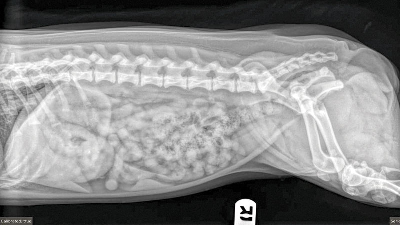
Studies tracking intact female dogs up to 10 years of age indicate that approximately 20 to 25 percent of all intact females will develop pyometra within their lifetimes. This incidence rate climbs dramatically when looking exclusively at specific breeds or aging populations over eight years old, where some cohorts demonstrate a lifetime risk exceeding 50 percent. This high prevalence serves as the primary justification behind proactive, early-age ovariohysterectomy recommendations by global veterinary associations.
Correct Answer to Question 2: c) 25 percent (Note: Many epidemiological studies cite ranges between 20% and 25%, with 25% serving as the widely accepted benchmark across academic curricula).
3) Primary Bacteriological Agents Involved
The pathogenesis of pyometra hinges on the interplay between a progesterone-primed, immunologically suppressed endometrium and ascending bacterial flora from the lower urogenital tract.
While multiple opportunistic pathogens can colonize the fluid-filled uterine lumen, by far the most common isolate retrieved from canine pyometra discharge is Escherichia coli (E. coli). Uropathogenic strains of E. coli possess specialized virulence factors, including P-fimbriae (adhesins) that enable the bacteria to bind specifically to endometrial receptors, resisting the mechanical clearance of uterine contractions.
Other less frequent isolates include Streptococcus species, Staphylococcus species, Pseudomonas aeruginosa, and Klebsiella pneumoniae. However, E. coli accounts for upwards of 70% to 80% of all confirmed bacteriological cultures from closed-cervix pyometra cases, frequently triggering systemic endotoxemia, localized peritonitis, and systemic inflammatory response syndrome (SIRS).
Correct Answer to Question 3: a) E. coli
Official Statements & Clinical Guidelines
Expert Commentary: Navigating the Surgical and Medical Crossroad
To gain a deeper perspective on the management of geriatric Bulldogs with acute pyometra, we consulted veterinary surgical specialists regarding the delicate balance between medical stabilization and immediate surgical intervention.
"The management of an acute, closed-cervix pyometra in a brachycephalic geriatric patient like an English Bulldog is a masterclass in emergency critical care," states Dr. Kendra Freeman, the attending clinician and author of this case presentation.
"Every hour a closed pyometra remains inside the abdomen increases the risk of uterine rupture, profound septic shock, and multi-organ dysfunction syndrome. Bulldogs present unique anesthetic challenges due to their brachycephalic airway anatomy, elongated soft palates, and hypoplastic tracheas. Therefore, rapid preoperative fluid resuscitation, meticulous electrolyte correction, and targeted antibiotic therapy must be balanced against the imperative to get the patient into surgery before endotoxemia becomes irreversible."
Consensus Guidelines from Veterinary Critical Care Societies
Recent updates from canine reproductive and emergency practice guidelines emphasize several mandatory pillars for pyometra management:
- Zero Tolerance for Medical Management in Closed Pyometra: While prostaglandin ($PGF_2alpha$) therapy combined with supportive care is occasionally debated for valuable breeding animals with open cervix pyometras, a closed-cervix presentation accompanied by systemic toxicity, azotemia, and abdominal rigidity mandates emergency ovariohysterectomy. Medical management in closed cases carries an unacceptably high rate of uterine rupture and mortality.
- Perioperative Fluid Resuscitation: Azotemic patients require aggressive balanced crystalloid therapy to restore glomerular filtration rates, correct electrolyte imbalances, and stabilize blood pressure prior to induction of general anesthesia.
- Anesthetic Protocol Tailoring: For Bulldogs and other brachycephalic breeds, premedication protocols should avoid excessive sedation that compromises respiratory drive. Induction agents must allow for rapid endotracheal intubation, and meticulous monitoring of SpO2, ETCO2, and blood pressure is non-negotiable throughout the extirpation of the septic uterus.
Future Outlook: Prevention, Early Screening, and Client Education
As veterinary medicine continues to advance, the overarching goal regarding pyometra is the complete eradication of preventable cases through client education and proactive surgical intervention.
The Role of Proactive Ovariohysterectomy
The most effective strategy against pyometra remains elective ovariohysterectomy performed prior to the first or second estrus cycle. Not only does this eliminate the target organ entirely, but early-stage spaying dramatically reduces the lifetime incidence of mammary neoplasia, eliminates the risk of ovarian and uterine tumors, and prevents accidental, unwanted litters.
Emerging Diagnostic Biomarkers
For clients who elect to maintain their female dogs intact for breeding or show purposes, veterinary researchers are currently investigating early predictive biomarkers for cystic endometrial hyperplasia. Advanced ultrasonographic elastography, serial serum progesterone monitoring, and novel inflammatory biomarkers (such as C-reactive protein [CRP] and serum amyloid A [SAA]) hold promise for identifying subclinical uterine changes long before systemic toxicity sets in.
Client Communication and Vigilance
Veterinary practitioners must continue to educate pet owners—particularly those managing high-risk breeds like Bulldogs, Golden Retrievers, and Rottweilers—about the subtle warning signs of metestrual illness. Owners must be counseled that lethargy, inappetence, increased water consumption (polydipsia secondary to endotoxin-induced nephrogenic diabetes insipidus), and any abnormal vaginal discharge occurring 4 to 8 weeks post-heat represent a veterinary emergency requiring immediate diagnostic evaluation.
Through a combination of rapid diagnostic workups, aggressive surgical execution, and unwavering dedication to preventive medicine, clinicians can successfully navigate these complex emergency presentations and secure favorable outcomes for vulnerable canine patients.