
By Veterinary News Investigative Desk
Specialized Reporting on Veterinary Dentistry and Oral-Maxillofacial Surgery
Executive Overview
When a feline patient presents to the clinic with classic clinical signs of sublingual distress—such as persistent bloody drool, sudden or progressive anorexia, and profound dysphagia (difficulty swallowing)—veterinary practitioners are conditioned by clinical odds to immediately suspect squamous cell carcinoma (SCC). In a 10-year-old cat, this grim diagnosis represents the statistical baseline. However, anchoring strictly to an SCC diagnosis can result in devastating diagnostic miscalculations, inappropriate treatment pathways, and premature, pessimistic prognoses.
A recent clinical encounter managed by Dr. John R. Lewis, VMD, DAVDC, Fellow-AVDC OMFS, serves as an invaluable reminder that the feline sublingual space can host a variety of non-malignant, inflammatory, traumatic, and obstructive pathologies. While oral SCC remains the most prevalent and aggressive malignant neoplasm of the feline oral cavity, clinicians must systematically broaden their differential diagnoses.
This comprehensive review explores the diagnostic complexities surrounding feline sublingual lesions. Drawing from expert insights, case studies, and clinical pearls, we examine alternative etiologies—ranging from fluid-filled ranulas and complex eosinophilic granuloma complexes to linear foreign bodies and chronic mechanical irritation. Ultimately, this article emphasizes that histopathology remains the gold standard, ensuring that every patient receives a tailored, accurate, and potentially life-saving therapeutic approach.
Detailed Chronology: Clinical Presentations and Diagnostic Pathways
To understand the diagnostic journey of sublingual lesions, one must trace how these abnormalities manifest, how they are mistakenly categorized, and how clinicians ultimately arrive at definitive diagnoses through systematic investigation.
The Statistical Weight of Squamous Cell Carcinoma
Clinicians evaluating older cats with oral bleeding, halitosis, and weight loss are justifiably primed to look for SCC. The disease originates from the mucosal epithelium and is notoriously invasive, often infiltrating underlying bone before detection. Because its physical appearance can occasionally mimic benign or inflammatory conditions, mistaking an aggressive tumor for a benign swelling—or conversely, writing off a treatable inflammatory mass as an inoperable SCC—carries profound clinical consequences.
The Enigma of the Ranula
When physical examination reveals a smooth, translucent or pinkish swelling beneath the tongue, the clinician’s differential should immediately shift toward a ranula. Deriving its name from the Latin word "rana" (meaning frog), a ranula visually and descriptively resembles a frog’s underbelly.
Ranulas develop due to the abnormal extravasation and accumulation of saliva, typically stemming from trauma, obstruction, or rupture of the salivary ducts or glands. Underneath the tongue of both felines and canines lie paired, raised anatomical structures near the midline of the caudal symphysis known as the caruncles. These caruncles serve as the vital duct openings for the mandibular and sublingual salivary glands.
If physical trauma, inflammation, or iatrogenic damage impacts these caruncles—or the delicate ducts coursing between the glands and the caruncles—saliva leaks into the surrounding interstitial tissues. This dynamic creates a smooth, typically non-inflamed swelling that can present unilaterally or bilaterally. While minor ranulas may occasionally resolve spontaneously, the biological reality of compromised ductal integrity often necessitates surgical intervention, specifically the excision of the mandibular and sublingual salivary glands, to prevent recurrence.
Surgical Interventions and "Pseudoranulas"
The clinical picture can be further complicated by therapeutic interventions. During complex oncologic procedures such as mandibulectomies in dogs and cats, surgeons must occasionally transect and ligate major salivary ducts.
In the immediate postoperative window, this transection can induce localized sublingual edema and swelling that perfectly mimics a true ranula. Dubbed "pseudoranulas," these transient edematous accumulations generally resolve spontaneously within a few days to weeks as lymphatic and collateral drainage normalizes. Interestingly, historical veterinary literature includes experimental studies—such as a 1978 investigation evaluating surgical trauma to salivary ducts and glands—demonstrating that ductal trauma does not universally result in clinical ranula formation. Nevertheless, contemporary surgical specialists emphasize that clinicians must maintain a high index of suspicion for pseudoranulas following oncologic resections to avoid unnecessary re-operation.
Unraveling Eosinophilic Granuloma Complexes
Perhaps one of the most versatile mimics in veterinary oral pathology is the eosinophilic granuloma. While veterinary professionals classically associate eosinophilic granulomas with the ulcerated upper lip lesions colloquially termed "rodent ulcers," the spectrum of feline eosinophilic disease is far wider.
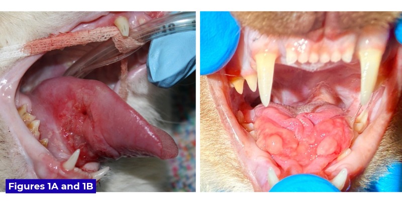
Manifestations can appear anywhere along the mucocutaneous junctions, lips, and sublingual architecture. Sublingual eosinophilic granulomas are particularly notorious because they can morphologically masquerade as either a smooth-surfaced ranula or an aggressive, verrucous (wart-like) squamous cell carcinoma.
A compelling case illustrating this diagnostic trap involved Milo, a one-year-old Ragdoll cat evaluated by Dr. Lewis alongside Dr. Kirk Herrmann, DAVDC, and Dr. Alex Juriga, DVM. Milo presented with a severe sublingual lesion that was verrucous, ulcerated, and remarkably firm to the touch—clinical features that heavily favored a diagnosis of SCC. Compounding the diagnostic puzzle, Milo possessed a concurrent clinical history of a swollen lower lip that remained partially inflamed upon presentation.
Recognizing the potential for severe self-trauma from occlusion against Milo’s dentition, the clinical team initiated medical management while awaiting diagnostic confirmation. Histopathological evaluation of an incisional biopsy revealed marked eosinophilic granulomatous glossitis with surface ulceration, confirming an eosinophilic granuloma rather than a neoplasm.
The treatment protocol involved an initial course of dexamethasone sodium phosphate transitioning rapidly to oral prednisolone at a tapered dosage of 1 mg/kg/day. The clinical response was dramatic: the sublingual swelling receded significantly within one week and achieved near-complete resolution by week two. Referral to a veterinary dermatologist was subsequently recommended to investigate underlying hypersensitivity or allergic etiologies, underscoring that medical management must extend beyond symptomatic relief to address root immunological triggers.
Supporting Context & Metrics: Rarities and Diagnostic Nuances
Beyond ranulas and eosinophilic granulomas, the feline sublingual space can harbor less common abnormalities that require careful clinical evaluation.
Linear Foreign Bodies and Anatomical Constrictions
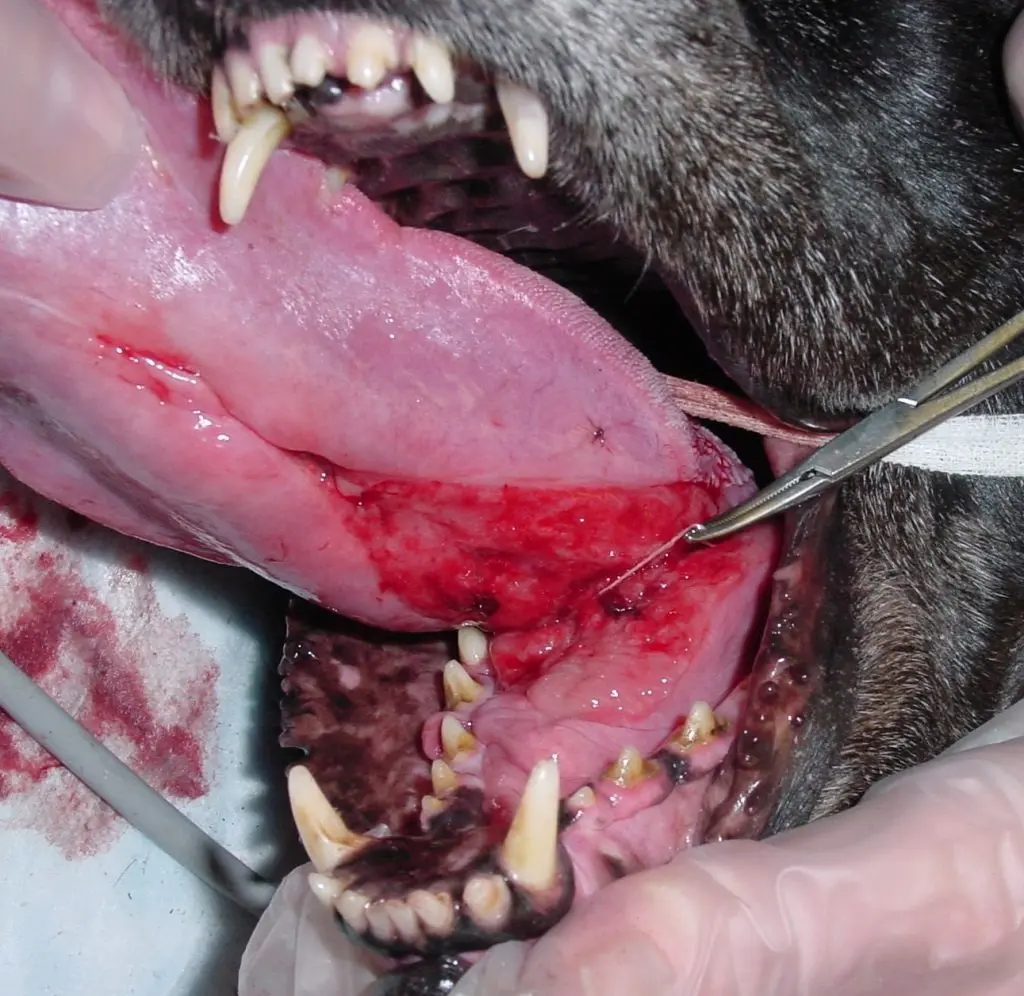
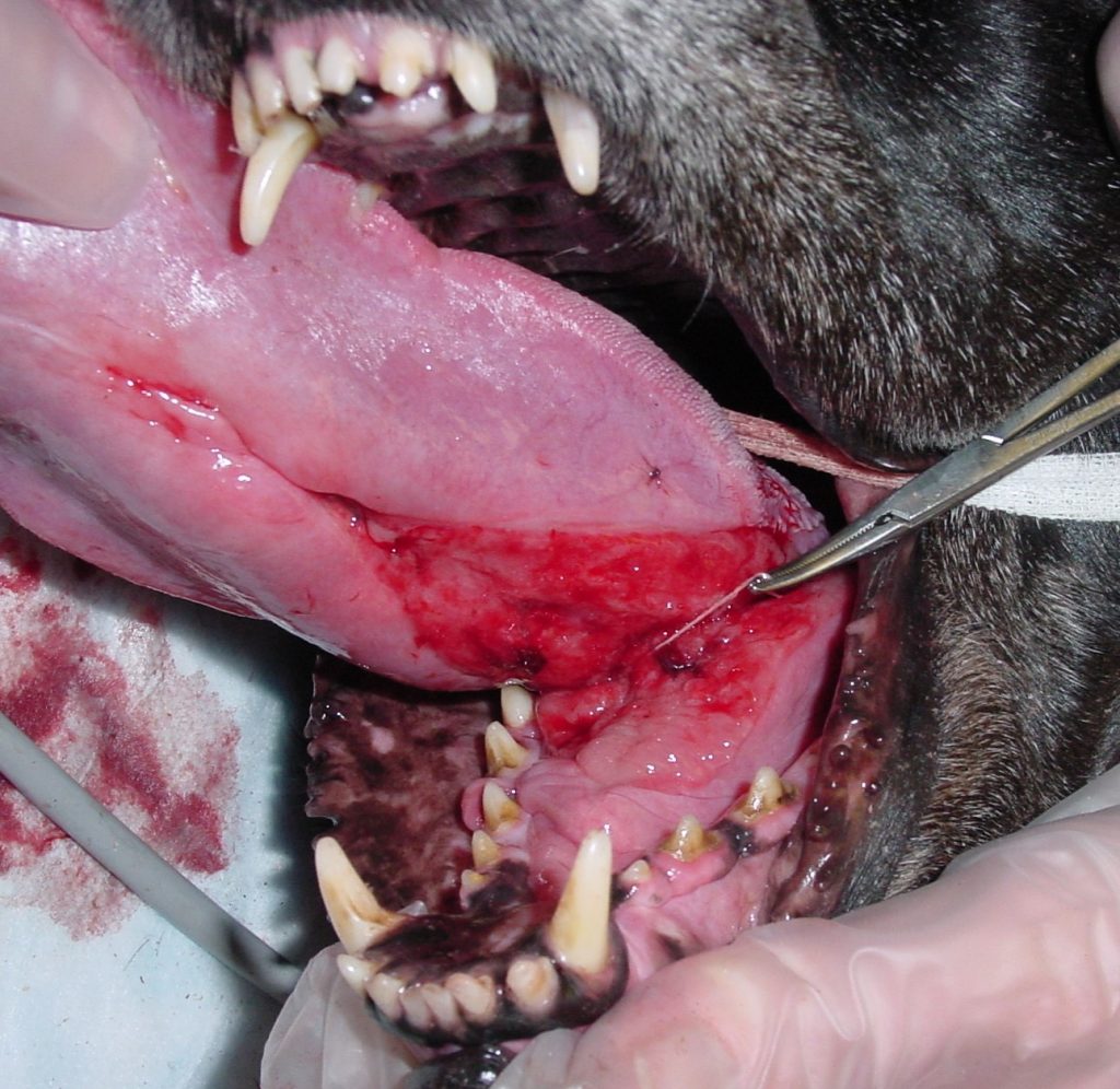
Linear foreign bodies—ranging from threads and strings to yarn, tinsel, and synthetic twines—represent a well-documented hazard in young, inquisitive felines, though they can occasionally affect canine patients as well. Dr. Lewis highlights a notable case involving a 10-year-old dog named Lily, who chewed on the inner twine of a golf ball, resulting in the material wrapping securely around the base of the tongue (the frenulum).
When a linear foreign body anchors around the lingual frenulum, normal tongue mobility and swallowing mechanics create a sawing effect. This dynamic leads to severe sublingual edema, intense erythema, mucosal laceration, and progressive swelling of the tongue body. If left undetected, linear foreign bodies can cause tissue necrosis and life-threatening gastrointestinal complications.
Chronic Mechanical Irritation and Granulation Tissue
Another rare but important differential diagnosis is the formation of exuberant granulation tissue in the sublingual space. Chronic mechanical irritation—such as constant friction against a sharp, jagged dental cusp altered by advanced tooth resorption—can stimulate localized tissue proliferation. This reactive mass can easily be mistaken for a neoplastic growth unless a thorough dental charting and oral examination are performed under general anesthesia.
| Lesion Type | Typical Signalment | Physical Characteristics | Primary Differential / Confirmatory Test |
|---|---|---|---|
| Squamous Cell Carcinoma (SCC) | Older cats (approx. 10 years) | Firm, ulcerated, locally invasive, painful | Incisional Biopsy / Histopathology |
| Ranula | Cats and dogs of various ages | Smooth, fluctuant, non-painful swelling beneath the tongue | Needle aspiration (salivary fluid analysis) |
| Eosinophilic Granuloma | Young to middle-aged cats (e.g., 1-year Ragdoll) | Firm, variable (smooth or verrucous), possible lip involvement | Incisional Biopsy / Histopathology |
| Linear Foreign Body | Inquisitive young cats (occasionally dogs) | Sublingual erythema, edema, anchored around frenulum | Visual inspection under sedation/anesthesia |
Official Statements and Expert Perspectives
The diagnostic challenges associated with sublingual lesions highlight the necessity of meticulous clinical protocols within veterinary dentistry and oral surgery.
Dr. John R. Lewis emphasizes that clinical intuition must always be backed by objective diagnostics:
"While playing the odds in an older cat with bloody drool points you toward squamous cell carcinoma, assuming every sublingual lesion is malignant is a disservice to our patients. Conditions like ranulas and eosinophilic granulomas can visually mimic cancer down to the last detail. Only through careful biopsy and histopathology can we protect our patients from misdiagnosis and deliver targeted, curative therapies."
Furthermore, collaborative cases—such as those involving multi-disciplinary insights from specialists like Dr. Kirk Herrmann and Dr. Alex Juriga—demonstrate that managing complex oral inflammation requires a holistic perspective. When immune-mediated conditions like the eosinophilic granuloma complex are identified, veterinary dentists stress the importance of partnering with veterinary dermatologists. Identifying and mitigating underlying environmental, parasitic, or food-related allergens is critical to preventing the chronic recurrence of oral lesions.
Future Outlook and Clinical Recommendations
As diagnostic imaging, histopathological techniques, and minimally invasive surgical modalities continue to advance, the veterinary community is increasingly equipped to differentiate complex oral lesions with unprecedented precision. However, the foundational rules of clinical practice remain unchanged.
Key Takeaways for Practitioners:
- Never Rely Solely on Signalment: While signalment (e.g., age, species) guides statistical probability, individual patients frequently deviate from epidemiological norms—as demonstrated by the one-year-old Ragdoll presenting with lesions mimicking advanced SCC.
- Embrace Histopathology as the Gold Standard: Visual inspection alone is insufficient for definitive diagnosis in the oral cavity. Incisional or excisional biopsies are mandatory to differentiate neoplastic processes from severe inflammatory or immune-mediated diseases.
- Consider Multidisciplinary Care: Managing immune-mediated conditions requires long-term medical strategies, including corticosteroid tapering protocols and dermatological evaluations to uncover root immunological triggers.
- Maintain a Broad Differential List: Keep ranulas, pseudoranulas, eosinophilic granulomas, linear foreign bodies, and reactive granulation tissue firmly on the radar when evaluating any sublingual abnormality.
By adhering to these rigorous diagnostic standards, veterinary professionals can ensure that every feline patient receives an accurate diagnosis, avoids unnecessary radical surgeries for benign conditions, and achieves the best possible quality of life.